MRI-Based Technology Identifies Stroke Recurrence Risk
By MedImaging International staff writers
Posted on 03 Jan 2016
Quantitative magnetic resonance angiography (QMRA) can be used to identify vertebrobasilar (VB) stroke patients who are at greater risk of having another stroke, according to a new study.Posted on 03 Jan 2016
Researchers at the University of Illinois in Chicago (UIC, USA), the University of Toronto (Canada), and other institutions conducted a prospective, blinded, longitudinal cohort study involving 72 patients with recent VB transient ischemic attack (TIA) or stroke, and 50% or more atherosclerotic stenosis or occlusion in VB arteries. The patients underwent large-vessel flow measurement using QMRA, and were also followed for two years at five academic medical centers as they continued receiving standard care from their neurologists, who were blinded to QMRA flow status.

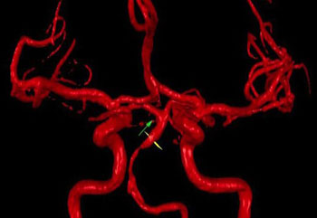
Image: MRI scan using NOVA technology (Photo courtesy of UIC).
The results showed that distal flow status was low in 25% of the patients and was significantly associated with risk for a subsequent VB stroke; 12- and 24-month event-free survival rates of 78% and 70%, respectively, were found in the low-flow group, compared to 96% and 87%, respectively, in the normal-flow group. Distal flow status remained significantly associated with risk even when controlling for the degree of stenosis and location. Medical risk factor management at 6-month intervals, however, was similar between patients with low and normal distal flow. The study was published in on December 21, 2015, in JAMA Neurology.
“At one year, the risk for patients with low blood flow was about five times as high as risk for patients without low flow in the back of the brain. For these patients, the benefits of angioplasty probably outweigh the risks,” said senior author professor of neurological surgery Sepideh Amin-Hanjani, MD. “About three-quarters of patients didn’t have low blood flow in the vertebrobasilar region. These patients would not benefit from treatments aimed at opening the vessels, such as angioplasty; in fact, the procedure would put these patients at unnecessary risk.”
The QMRA data was analyzed using noninvasive optimal vessel analysis (NOVA), a software program that can quantify the volume, velocity, and direction of blood flowing through any major vessel in the brain using standard MRI equipment. NOVA is a product of Vassol (River Forest, IL, USA), and was developed at UIC by head of neurological surgery Professor Fady Charbel, MD, who is also a coauthor of the new study.
Related Links:
University of Illinois in Chicago
University of Toronto
Vassol


