Study Shows Occurrence of Concussion in Military Personnel with Blast-Related Injuries to Be Surprisingly High
By MedImaging International staff writers
Posted on 16 Dec 2015
The results of a new study published in the online edition of the journal Radiology shows that brain damage in active duty military personnel with blast-related injuries is evident in a surprisingly high percentage of cases.Posted on 16 Dec 2015
Concussion or Mild Traumatic Brain Injury (MTBI) is very common among US military personnel returning from fighting in Iraq, and Afghanistan, with over 300,000 of them diagnosed with MTBI between 2000 and 2015. Currently MTBI injuries are assessed mainly using behavioral observations, and symptoms such as post-traumatic amnesia, and loss of consciousness. The need for a more definitive marker spurred the research team to use advanced Magnetic Resonance (MR) brain imaging as a tool for assessing MTBI, and to try to link the acquired MRI data with subjective MTBI symptoms. The researchers are trying to use the technique to diagnose Post-Traumatic Stress Disorder (PTSD), which has similar symptoms to TBI, but different treatments.

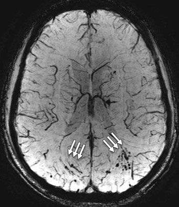
Image: The susceptibility-weighted image shows extensive micro-hemorrhage (see arrows) consistent with diffuse axonal injury in a 25-year-old man with blast-related mild TBI (Photo courtesy of RSNA).
The ability to show evidence of micro-hemorrhages, hyper-intense areas, and softening or loss of brain tissue in the advanced MRI scans also allows military personnel and their families to visualize the previously invisible “wounds of war.”
Gerard Riedy, MD, PhD, US National Intrepid Center of Excellence (NICoE; Bethesda, Maryland, USA), said, "We were really surprised to see so much damage to the brain in the MTBI patients. It's expected that people with MTBI should have normal MRI results, yet more than 50 percent had these abnormalities. This paper is just the tip of the iceberg. We have several more papers coming up that build on these findings and look at brain function, brain wiring, connectivity and perfusion, or brain blood flow. A scar on a brain scan is an objective finding. We start with the objective and build a foundation for the correct diagnosis of MTBI and then bring in the subjective measures later. An objective measure of traumatic brain injury can lead to proper therapies. Military traumatic brain injury is not a small problem for our country. Through this research, we hope to learn more about what the future entails for our military personnel who've suffered these injuries."
Related Links:
NICoE


 Guided Devices.jpg)


