New Study Shows MRI and CT Imaging Could Improve Treatment of Patients with COPD
By MedImaging International staff writers
Posted on 12 Jul 2015
The findings of a new study published online in the journal Radiology, show that treatment for some patients with Chronic Obstructive Pulmonary Disease (COPD) can be improved using imaging.Posted on 12 Jul 2015
Diagnosis of COPD often includes a test called spirometry, which tests lung function. A spirometry test results in a Forced Expiratory Volume in one second (FEV1) score but provides an incomplete measure of the functioning of the respiratory system.

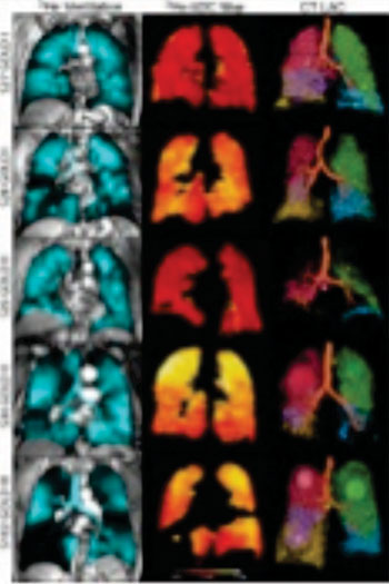
Image: Images in representative patients with mild-to-moderate or severe chronic obstructive pulmonary disease (COPD) (Photo courtesy of RSNA).
The researchers studied a group of 116 patients with COPD, and performed Computed Tomography (CT) and inhaled noble gas Magnetic Resonance Imaging (MRI) exams, in addition to FEV1 lung capacity testing. The patients also filled out a quality of life questionnaire, and walked for six-minutes to measure their exercise tolerance.
The results showed that in patients with mild-to-moderate COPD, and modestly abnormal FEV1 results, measurements of emphysema using MRI imaging correlated strongly with exercise limitation. Using both CT and MRI imaging and emphysema measurements helped clinicians explain COPD symptoms.
According to Grace Parraga, PhD, from the Robarts Research Institute (London, ON, Canada), and coauthor of the study, the implications of the findings are significant for patients with mild COPD and abnormal FEV1.
Grace Parraga, said, "COPD is a very heterogeneous disease. Patients are classified based on spirometry, but patients with the same air flow may have different symptoms and significant variation in how much regular activity they can perform, such as walking to their car or up the stairs in their home. FEV1 doesn't tell the whole story. With lung imaging, we can look at patients with mild disease much more carefully and change treatment if necessary."
Related Links:
RSNA


.jpg)