MRI Imaging Algorithm Helps Doctors Analyze Effectiveness of Hip Replacement Surgery
By MedImaging International staff writers
Posted on 22 Jun 2015
A new Magnetic Resonance Imaging (MRI) imaging tool has been developed that enables surgeons to determine the effectiveness of hip replacement surgery, and spot inflammation and adverse reactions before they can spread.Posted on 22 Jun 2015
The MRI algorithm was designed by Dr. Hollis G Potter, MD, chairman of the Department of Radiology and Imaging at the Hospital for Special Surgery (New York, NY, USA), and developed into an imaging tool by scientists at GE Healthcare (Chalfont St Giles, Buckinghamshire, UK).

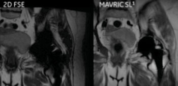
Image: Comparison of Hip Imaging Using the 2-D Fast Spin Echo (FSE), and the MAVRIC SL Imaging Tool (Photo courtesy of the Hospital for Special Surgery, New York, NY, USA).
Most hip replacements consist of nonmagnetic material that can scanned in an MRI machine, however, the implants often appear blurred, leave artifacts, or distort images.
The MAVRIC SL imaging tool was designed to significantly reduce artifacts from MR conditional metallic implants, and improve visualization of bone and soft tissue, enabling clinicians to spot adverse tissue reactions before they can do serious damage.
Dr. Potter, explained, “When you put the same component (in this case, a hip implant) into a group of different people, some people do beautifully, but other people will have an inflammatory reaction to the implant and fail quickly, requiring revision surgery. So we need a means by which to noninvasively visualize this inflammatory reaction process. The traditional techniques, including measurement of blood ion levels of the metallic components, have not proved efficacious. We’ve spent about 10 to 12 years studying patients with [hip] implants and found that MRI is very helpful in the early detection of an adverse reaction to an implant.”
Related Links:
GE Healthcare
Hospital for Special Surgery


.jpg)
