Neurosurgeons Use Novel MRI Diffusion Tensor Imaging Technique to Safely Remove Nerve Tumors
By MedImaging International staff writers
Posted on 02 Jun 2015
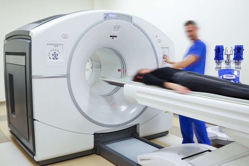
A neurosurgeon and neuroradiologist have for the first time successfully applied Diffusion Tensor Imaging (DTI) to map nerve fibers during surgery preparations for the safe removal of nerve tumors.Posted on 02 Jun 2015
The unique Magnetic Resonance Imaging (MRI) sequencing technique allows neurosurgeons to map a route around nerve fibers, and access/remove tumors without damaging the nerves. Traditional MRI scans are often unable to present a comprehensive view of each patient’s unique nerve fibers.
DTI uses the diffusion of water molecules in and around nerves to provide color 2-D or 3-D images of nerve fibers, and their relationship to schwannomas, a rare type of nerve tumor. The technique was developed by Northwestern Medicine (Chicago, IL, USA) neurosurgeon Michel Kliot, and functional neuroradiologist Thomas A. Gallagher.
Thomas A. Gallagher, said, "DTI can give us additional information about the organization of nerves as well as the diffusion properties within tumors; this is something that conventional MRI cannot do. Applications of this technology could extend beyond nerve tumors, and give us insight into many different nerve pathologies, traumatic nerve injuries and even the regrowth or recovery of nerves over time."
Related Links:
Northwestern Medicine

Gold Member
Solid State Kv/Dose Multi-Sensor
AGMS-DM+

New
X-Ray Detector
FDR-D-EVO III

Ultrasound Needle Guide
Ultra-Pro II

New
Color Doppler Ultrasound System
KC20

.jpg)