Physicians Advocate the Use of Ultrasound for Investigating Pelvic Symptoms
By MedImaging International staff writers
Posted on 13 May 2015
The American Institute of Ultrasound in Medicine (AIUM; Laurel, MD, USA) has published an article in the April 2015 issue of the American Journal of Obstetrics & Gynecology recommending the use of ultrasound examinations instead of Computed Tomography (CT), or Magnetic Resonance Imaging (MRI), for women presenting with flank pain, pelvic pain, or pelvic masses.Posted on 13 May 2015
The recommendation comes as part of a the AIUM’s “Ultrasound First,” campaign which advocates the use of ultrasound examinations when there is evidence that it is as effective as CT or MRI examinations for the target anatomic area. Ultrasound is the least invasive, least expensive modality, and avoids exposure to radiation. Not uncommonly, the results of MRI or CT examinations of the abdomen need clarification by ultrasound imaging.

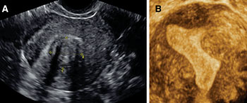
Image: Submucosal fibroid seen using 2-D and 3-D ultrasound (Photo courtesy of Benacerraf, Am J Obstet Gynecol 2015).
The authors of the recommendation state that the use of CT scans in the USA has tripled since 1993 and could result in 29,000 new cases of cancer, especially from CT of the pelvis and abdomen.
The article presents the current capabilities of ultrasound imaging technology that now offers multiple applications such as 3-D volume imaging, real-time evaluation of pelvic organs together with a physical examination, and Doppler blood flow mapping.
The authors conclude by recommending the use of ultrasound as the first-line imaging technique for the pelvis of women who are not pregnant, in most clinical scenarios. Consistent use of ultrasound imaging with new 3-D, and Doppler techniques could be more cost-effective and safer than CT or MRI, and render further imaging unnecessary. Users of ultrasound equipment are not always able to operate the modality to its full potential, and therefore there is also a need for further education and training.
Related Links:
American Institute of Ultrasound in Medicine


.jpg)

