DTI-MRI Shows Brain Alterations in High School Football Players After Only One Season
By MedImaging International staff writers
Posted on 16 Dec 2014
Some high school football players exhibit measurable brain alterations after only one season of play, even in the absence of concussion.Posted on 16 Dec 2014
The new findings were presented at the annual meeting of the Radiological Society of North America (RSNA), held November 30 through December 5, 2014, in Chicago (IL, USA). “This study adds to the growing body of evidence that a season of play in a contact sport can affect the brain in the absence of clinical findings,” said Christopher T. Whitlow, MD, PhD, MHA, associate professor of radiology at Wake Forest School of Medicine and radiologist at Wake Forest Baptist Medical Center (Winston-Salem, NC, USA).
 helmet-mounted accelerometers are used in youth and collegiate football to evaluate the frequency and severity of helmet impacts (Photo courtesy of RSNA).")
Image: Head impact telemetry system (HITs) helmet-mounted accelerometers are used in youth and collegiate football to evaluate the frequency and severity of helmet impacts (Photo courtesy of RSNA).
There have been various recent reports about the potential effects playing youth sports may have on developing brains. However, most of these studies examined brain changes as a result of concussion. Dr. Whitlow and colleagues set out to determine if head impacts acquired over a season of high school football produce white matter changes in the brain in the absence of clinically diagnosed concussion.
The researchers studied 24 high school football players between the ages of 16 and 18. For all games and practices, players were monitored with head impact telemetry system (HITs) helmet-mounted accelerometers, which are used in youth and collegiate football to evaluate the frequency and severity of helmet impacts. Risk-weighted cumulative exposure was computed from the HITs data, representing the risk of concussion over the course of the season. This data, along with total impacts, were used to classify the players into one of two groups: heavy hitters or light hitters. There were 9 heavy hitters and 15 light hitters. None of the players experienced concussion during the season.
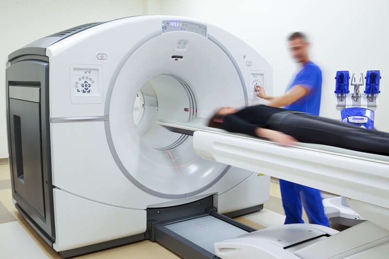
All players underwent pre- and post-season evaluation with diffusion tensor imaging (DTI) of the brain. DTI is a sophisticated magnetic resonance imaging (MRI) technique, which identifies microstructural changes in the brain’s white matter. The brain’s white matter is made up of millions of nerve fibers called axons that act similar to communication cables connecting various regions of the brain. Diffusion tensor imaging generates a measurement, called fractional anisotropy (FA), of the movement of water molecules along axons. In healthy white matter, the direction of water movement is comparatively uniform and measures high in fractional anisotropy. When water movement is more haphazard, fractional anisotropy values decrease, suggesting microstructural abnormalities.
The study’s findings demonstrated that both groups demonstrated global increases of FA over time, likely reflecting effects of brain development. However, the heavy-hitter group showed statistically significant areas of decreased FA post-season in specific areas of the brain, including the splenium of the corpus callosum and deep white matter tracts. “Our study found that players experiencing greater levels of head impacts have more FA loss compared to players with lower impact exposure,” Dr. Whitlow said. “Similar brain MRI changes have been previously associated with mild traumatic brain injury. However, it is unclear whether or not these effects will be associated with any negative long-term consequences.”
Dr. Whitlow cautioned, however, that these findings are preliminary, and more research still needs to be conducted.
Related Links:
Wake Forest Baptist Medical Center

Gold Member
Solid State Kv/Dose Multi-Sensor
AGMS-DM+

New
Enterprise Imaging & Reporting Solution
Syngo Carbon

New
Ceiling-Mounted Digital Radiography System
Radiography 5000 C

New
Mobile Digital C-arm X-Ray System
HHMC-200D

.jpg)