


e-Incubator Technology Provides Real-Time Imaging of Bioengineered Tissues in a Controlled Unit
By MedImaging International staff writers
Posted on 24 Nov 2014
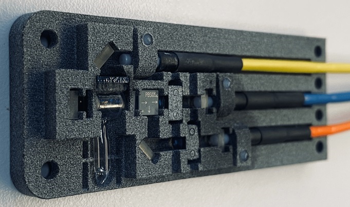
A new e-incubator, an innovative miniature incubator that is compatible with magnetic resonance imaging (MRI), enables scientists to grow tissue-engineered constructs under a controlled setting and to study their growth and development in real time without risk of contamination or damage. Posted on 24 Nov 2014
Offering the potential to test engineered tissues before human transplantation increases the success rate of implantation, and accelerates the translation of tissue engineering methods from the lab to the clinic, the novel e-incubator was described in the journal Tissue Engineering, Part C.
Shadi Othman, PhD, Karin Wartella, PhD, Vahid Khalilzad Sharghi, and Huihui Xu, PhD, from the University of Nebraska-Lincoln (USA), presented their findings of a validation study using the device to culture tissue-engineered bone constructs for four weeks. The e-incubator is a standalone unit that automatically detects and regulates internal conditions such as temperature, carbon dioxide levels, and pH via a microcontroller. It performs media exchange to feed the cultures and remove waste products. The current design is compatible with MRI to monitor the constructs without removing them from the incubator. With proper adjustments, compatibility with other imaging technologies including computed tomography [CT] and optical imaging is also possible.”
“Calibratable, hands-free tissue development environments are becoming increasingly important for the engineering of implantable tissues,” said Tissue Engineering co-editor-in-chief Peter C. Johnson, MD, vice president, research and development, Avery Dennison Medical Solutions (Chicago, IL, USA), and president and CEO, Scintellix, LLC (Raleigh, NC, USA). “In this new development, noninvasive imaging modalities are added to the spectrum of sensing and environmental capabilities that heretofore have included temperature, humidity, light, physical force, and electromagnetism. This represents a solid advance for the field.”
Related Links:
University of Nebraska-Lincoln

Gold Member
Solid State Kv/Dose Multi-Sensor
AGMS-DM+

Silver Member
Mobile X-Ray Barrier
Lead Acrylic Mobile X-Ray Barriers

New
Compact C-Arm
Arcovis DRF-C S21

Ultrasound System
Acclarix AX9

.jpg)