Ultrahigh-Field fMRI Reveals Language Hubs in the Brain in Great Detail
By MedImaging International staff writers
Posted on 12 Nov 2014
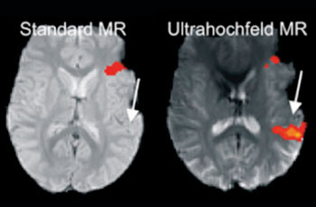
For the first time, new research has demonstrate that the areas of the brain that are important for understanding language can be targeted much more effectively using ultrahigh-field, 7 Tesla magnetic resonance imaging (MRI) technology than with standard clinical MRI scanners. This helps to protect these areas more effectively during brain surgery and avoid inadvertently damaging it. Posted on 12 Nov 2014
Before brain surgery, it is important to precisely understand the areas of the brain required for language in order to avoid injuring them during the procedure. Their position can shift considerably, especially in patients with tumors or brain injuries. The brain’s flexibility also means that language centers can shift to other regions. If the areas responsible for language control and processing are injured during a brain operation, the patient can be left unable to communicate. To generate a map of the language control centers prior to the operation, functional MRI (fMRI) is now used.

Image: Ultra-high-field MRI reveals language centers in the brain in much more detail (Photo courtesy of Medical University of Vienna).
A multicenter study from 2013 demonstrated the advantages of fMRI-assisted localization of the motor centers in the brain. In a new investigation by the working group led by Dr. Roland Beisteiner, from the department of neurology at the Medical University of Vienna (Austria) it has been possible for the first time to demonstrate that the areas of the brain that are important for understanding language can be pinpointed even more accurately using ultra-high-field 7 Tesla MRI than with traditional clinical MRI scanners. The focus lies on the two most important language centers in the brain known as Wernicke’s area (which controls the understanding of language) and Broca’s area, which controls the motor functions involved with speech.
The brain is scanned for activity while the patient is performing speech tasks. This allows the areas required for speech to be localized much more accurately than previously. “Ultrahigh-field MR offers much greater sensitivity than classic MRI scanners,” explained Dr. Beisteiner, “allowing even very weak signals to be recorded in areas that would otherwise have been missed.”
The research was conducted in cooperation between the University department of radiology and nuclear medicine and other university departments, and was published September 22, 2014, in the journal Neuroimage. The working group of this study has published numerous articles on the improvement of neurologic diagnostics using fMRI and performed the first study into improving the diagnosis of brain function using ultrahigh-field MRI. Today, fMRI is the most important noninvasive technique in Austria for the research and clinical investigation of brain functions.
Related Links:
Medical University of Vienna


