MRI Helps to Identify and Quantify Fat in the Liver
By MedImaging International staff writers
Posted on 28 Sep 2014
Spanish investigators have shown that magnetic resonance imaging (MRI) is a good modality—better than hepatic biopsy—for identifying fats in the liver and for quantifying them.Posted on 28 Sep 2014
Excess weight causes significant alterations in the body, one of which affects liver function. Fat accumulates in the liver producing hepatic steatosis, which, in specific circumstances, causes inflammation, fibrosis, and ultimately, cirrhosis. To date, the most effective method for determining hepatic fat has been hepatic biopsy. Imaging techniques such as abdominal echocardiography are less precise for determining the quantity of fat.

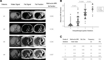
Image: Multi-echo MRI fat fractions positively correlate with the grade of steatosis estimated by histopathologic measurements. (A) Representative multi-echo MRI images showing different degrees of water and fat intensity, and fat fraction in different patients. (B) Multi-echo MRI fat fractions positively correlate with the grade of steatosis estimated by histopathologic measurements in human livers (n =129). Dots represent the values of each case. (C) Multi-echo MRI fat fraction mean values of each steatosis grading group (0 to 3 scale). CV, coefficient of variation; MRI, magnetic resonance imaging; Std. Dev, standard deviation (Photo courtesy of Jiménez-Agüero, et al: BMC Medicine 2014 12:137).
The research team led by Luis Bujanda, a professor of medicine at the University of the Basque Country (UPV/EHU; Leioa, Spain), in charge of the research into hepatic and gastrointestinal diseases at the Biodonostia Health Research Institute, conducted the study.
Obesity and overweight affect more than 50% of the population in the Basque community. The research was been published August 26, 2014, in the BMC Medicine, and coordinated by Drs. Jesús Bañales of the Biodonostia Health Research Institute (HRI; Gipuzkoa, Spain) and Raúl Jimenez of the department of surgery, radiology, and physical medicine at the UPV/EHU’s medicine and odontology faculty. In addition, the study has had the participation of researchers from the department of nutrition and food sciences of the UPV/EHU’s pharmacy faculty, from the surgery, digestive system, and pathological anatomy services at the Donostia University Hospital, together with Osatek (the Basque Public Health department’s imaging diagnosis service).
The research was undertaken with 97 obese patients and 32 patients with other hepatic pathologies, and who had been subject to surgical intervention. The quantity of fat in the liver was measured using three different methods; MRI, hepatic biopsy, and the biochemical determination of fat employing the Folch method. Patients were subject to MRI scanning the day before surgery and a sample of liver was obtained during the surgery.
“Magnetic resonance is a very useful technique to determine the presence of fat or not in the liver, the quantity of the same and in order to evaluate the efficacy of treatment applied over a long period. It is possible that in the future we will be able to determine, apart from fat, the degree of inflammation and hepatic fibrosis,” stated Dr. Bañales, a researcher at Biodonostia HRI.
The article validates the earlier research undertaken in lab animals and published by the same research team one year ago in which it was observed that the quantification of hepatic fat was very precise when using MRI scanning.
Related Links:
University of the Basque Country
Biodonostia Health Research Institute


