fMRI Shows Neural Compensation Found in Individuals with Alzheimer’s-Related Protein
By MedImaging International staff writers
Posted on 24 Sep 2014
A neuroimaging study has demonstrated that the human brain is capable of a neural plasticity that compensates for the accumulation of beta-amyloid, a destructive protein linked with Alzheimer’s disease.Posted on 24 Sep 2014
The findings, published September 14, 2014, in the journal Nature Neuroscience, could help clarify how some older adults with beta-amyloid deposits in their brain maintain normal cognitive function while others go on to develop dementia.
“This study provides evidence that there is plasticity or compensation ability in the aging brain that appears to be beneficial, even in the face of beta-amyloid accumulation,” said study principal investigator Dr. William Jagust, a professor with joint appointments at the University of California (UC) Berkeley’s (CA, USA) Helen Wills Neuroscience Institute, the School of Public Health, and Lawrence Berkeley National Laboratory (Berkeley, CA, USA).
Earlier research had shown a connection between increased brain activity and beta-amyloid deposits, but it was unclear whether the activity was tied to better mental performance. The study included 22 healthy young adults and 49 older adults who had no signs of mental decline. Brain scans showed that 16 of the older study participants had beta-amyloid deposits, while the remaining 55 adults did not.
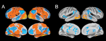
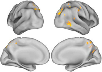
The researchers employed functional magnetic resonance imaging (fMRI) to monitor the brain activity of subjects in the process of memorizing pictures of various scenes. Subsequently, the researchers tested the participants’ “gist memory” by asking them to validate whether a written description of a scene, such as a boy doing a handstand, corresponded to a picture earlier viewed. Individuals were then asked to confirm whether specific written minutiae of a scene, such as the color of the boy’s shirt, were correct.
“Generally, the groups performed equally well in the tasks, but it turned out that for people with beta-amyloid deposits in the brain, the more detailed and complex their memory, the more brain activity there was,” said Jagust. “It seems that their brain has found a way to compensate for the presence of the proteins associated with Alzheimer’s.”
What is still uncertain, according to Dr. Jagust, is why some people with beta-amyloid deposits are better at using different parts of their brain than others. Previous studies suggest that people who engage in mentally stimulating activities throughout their lives have lower levels of beta-amyloid. “I think it’s very possible that people who spend a lifetime involved in cognitively stimulating activity have brains that are better able to adapt to potential damage,” said Dr. Jagust.
Related Links:
University of California Berkeley


.jpg)