Neuro-MRI Scanning Reveals Disparities Between Imagining and Remembering
By MedImaging International staff writers
Posted on 10 Sep 2014
Using functional magnetic resonance imaging (fMRI) technology, investigators are learning more about where and how imagination processes occur in human brains. Investigators devised a study that would help them distinguish real imagination from related mental processes such as remembering. Posted on 10 Sep 2014
The study was devised by graduate student from Brigham Young University (BYU; Salt Lake City, UT, USA), Stefania Ashby, and her faculty mentor. “I was thinking a lot about planning for my own future and imagining myself in the future, and I started wondering how memory and imagination work together,” Ms. Ashby said. “I wondered if they were separate or if imagination is just taking past memories and combining them in different ways to form something I’ve never experienced before.”

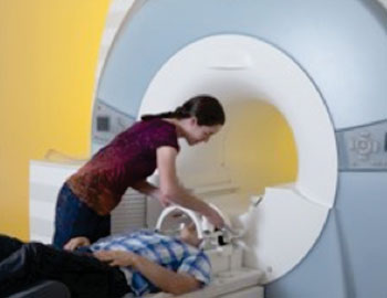
Image: BYU\'s MRI lab (Photo courtesy of Mark A. Philbrick).
Scientists have been debating over whether memory and imagination are in reality distinct processes. They asked study participants to provide 60 personal images for the “remember” section of the study. Participants also filled out a questionnaire earlier to determine which settings would be unfamiliar to them and consequently a better fit for the “imagine” section.
The researchers then showed study participants their own photographs during an fMRI scanning session to provoke brain activity that is precisely memory-based. A statistical analysis demonstrated distinctive patterns for memory and imagination. “We were able to see the distinctions even in those small regions of the hippocampus,” Ms. Ashby said. “It’s really neat that we can see the difference between those two tasks in that small of a brain region.”
Ms. Ashby is currently working as a research associate at the University of California (UC) Davis (USA), where she uses neuroimaging to study individuals at risk of psychotic disorders such as schizophrenia. Her plan is to earn a PhD in neuroscience and continue researching.
The study’s findings were published June 26, 2014, in the journal Cognitive Neuroscience.
Related Links:
Brigham Young University


