Research Demonstrates MRI Acts as Predictive Marker for Epilepsy Development Following Febrile Seizure
By MedImaging International staff writers
Posted on 16 Jul 2014
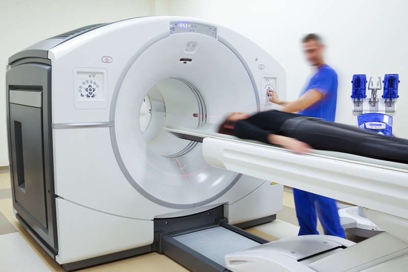
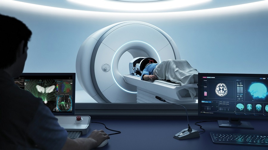
Within hours of a fever-induced seizure, magnetic resonance imaging (MRI) may be able to identify brain changes that occur in those most likely to develop epilepsy later in life, according to recent animal research. The findings may soon help improve ways to detect children at an increased risk for developing epilepsy and direct efforts to prevent epilepsy development in those at greatest risk. Posted on 16 Jul 2014
Convulsions brought on by fever, febrile seizures typically last only a few minutes and are relatively common in infants and small children. However, in some cases, children experience febrile seizures that last for more than 30 minutes (known as febrile status epilepticus [FSE]). Of these children, 40% will go on to develop temporal lobe epilepsy (TLE)—a typical and frequently treatment-resistant brain disorder. Physicians currently have no way to anticipate which of the children with a history of extended febrile seizures (FSE) will go on to develop TLE, and children typically do not experience the beginning of the disease until 10–12 years after the onset of FSE.
Tallie Z. Baram, MD, PhD, and her colleagues at the University of California-Irvine (USA), in this study, used MRI scanning to examine the brains of young rats right after after FSE was induced to compare the brains of the animals that would go on to develop TLE and those that would not. The researchers, who published their findings June 25, 2014, in the Journal of Neuroscience, monitored the rats as they developed over 10 months for signs of TLE. Of the animals that developed epilepsy over the course of the study, all had a distinctive MRI signal in a part of the brain called the amygdala when imaged within hours after the FSE. This signal was not visible in the rats that remained epilepsy-free for the duration of the experiment.
“This remarkable discovery got us to ask two key questions,” Dr. Baram said. “First, can we figure out what is going on in the brain that causes this new signal? And second, can we detect a similar predictive signal in children after febrile status epilepticus?”
Additional study into the origin of the MRI signal revealed that the brains of the lab rats that went on to develop epilepsy were consuming more energy and using up more oxygen in the amygdala hours after long febrile seizures than the brains of the rats that did not develop epilepsy later in life. “Detecting reduced oxygen may be an early marker of brain damage that leads to subsequent spontaneous seizures and epilepsy,” explained Hal Blumenfeld, MD, PhD, who studies epilepsy at Yale University (New Haven, CT, USA), and was not involved in this study.
Although the current study was conducted in rodents using a high-power laboratory scanner, additional studies by Dr. Baram’s group revealed that the epilepsy-predicting signal could be detected using a standard hospital MRI scanner. This indicates that similar evaluations could be done in children with FSE to begin to evaluate whether this signal appears in children after FSE and whether it predicts the emergence of epilepsy later on in life.
“Preventive therapy development is hampered by our inability to identify early the individuals who will develop TLE,” Dr. Baram explained. “Finding a predictive signal using clinically applicable noninvasive brain scans holds promise for predicting epilepsy after FSE.”
Related Links:
University of California-Irvine

Gold Member
Solid State Kv/Dose Multi-Sensor
AGMS-DM+

Silver Member
Mobile X-Ray Barrier
Lead Acrylic Mobile X-Ray Barriers

New
Ultrasound Table
Ergonomic Advantage (EA) Line

Portable Radiology System
DRAGON ELITE & CLASSIC
.jpg)