Four Imaging Approaches Can Differentiate Malignant and Benign Breast Tumors
By MedImaging International staff writers
Posted on 10 Jul 2014
Imaging breast tumors using four approaches together, can better distinguish malignant breast tumors from those that are benign, compared with imaging using fewer approaches, and this may help avoid repeat breast biopsies. Posted on 10 Jul 2014
The findings were published online June 24, 2014, in Clinical Cancer Research, a journal of the American Association for Cancer Research. “By assessing many functional processes involved in cancer development, a multiparameter PET/MRI [positron emission tomography/magnetic resonance imaging] of the breast allows for a better differentiation of benign and malignant breast tumors than currently used dynamic contrast-enhanced MRI [DCE-MRI)] by itself. Therefore, unnecessary breast biopsies can be avoided,” said Katja Pinker, MD, associate professor of radiology in the department of biomedical imaging and image-guided therapy at the Medical University of Vienna (Austria).

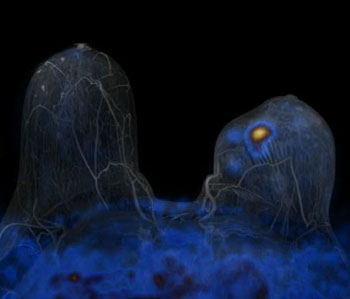
Image: A PET/MRI scanner visualizes breast cancer in this fused PET/MR image (Photo courtesy of Medscape).
The new imaging technique, called multiparametric (MP) 18FDG (fluorodeoxyglucose) PET-MRI, which used four imaging approaches, was 96% accurate in distinguishing malignant breast tumors from those that were benign, and provided better results than combinations of two or three imaging approaches. The study estimates that this technique can cut unnecessary breast biopsies recommended by the typically used imaging modality, the DCE-MRI, by 50%.
“DCE-MRI is a very sensitive test for the detection of breast tumors, but is limited in visualizing the functional properties cancer cells acquire during development. Therefore, there is still room for improvement,” explained Dr. Pinker. “PET/MRI mirrors cancer biology and allows accurate diagnosis of breast cancer without a biopsy. Additionally, the more accurately we understand a tumor’s biology, the better we can tailor therapy to each breast cancer patient’s individual needs. Provided that a hospital is equipped with a PET/CT [computed tomography] and an MRI scanner or a combined PET/MRI, the technique we have described can be immediately implemented in clinics,” said Dr. Pinker.
The investigators recruited 76 patients to the study who had suspicious or inconclusive findings from a mammography or a breast ultrasonography. They performed a MP18FDG PET/MRI on all the patients. Furthermore, all patients’ breast tumor biopsies were evaluated by histopathology.
To determine the combination of imaging parameters that yielded the most accurate results, Dr. Pinker and colleagues combined the imaging data from two parameters, three parameters, and all four parameters. All two-parameter and three-parameter evaluations included DCE-MRI. All findings were compared with histopathology diagnosis to evaluate which combination was most efficient in making an accurate diagnosis. Of the 76 tumors, based on histopathology, 53 were malignant and 23 were benign.
The researchers found that none of the two- or three-parameter combinations reached the same level of sensitivity and specificity as the four-parameter method, which had an appropriate use criteria (AUC) of 0.935. (An AUC of 0.9 to 1 means the modality is excellent, and an AUC of 0.5 means the method is worthless.) “Performing a combined PET-MRI is currently less cost-effective than existing breast imaging methods,” said Dr. Pinker. “However, a significant reduction in unnecessary breast biopsies by using this combined method may improve the cost-effectiveness.”
MP18FDG PET-MRI allowed tumor imaging by four parameters: DCE-MRI, diffusion-weighted imaging (DW), three-dimensional (3D) 1H-magnetic resonance spectroscopic imaging (MRSI), and 18FDG-PET.
Related Links:
Medical University of Vienna


.jpg)