Concussion Recovery Time May Take Longer for Men
By MedImaging International staff writers
Posted on 22 May 2014
A study of concussion patients using diffusion tensor imaging (DTI) found that males took longer to recover after concussion than females did. The study’s findings revealed that DTI can be used as a bias-free approach to predict concussion outcome. Posted on 22 May 2014
The study’s findings were published online May 6, 2014, in the journal Radiology. More than 17 million individuals in the United States suffer a mild traumatic brain injury (mTBI) yearly, also known as a concussion, of which approximately 15% suffer persistent symptoms beyond three months.

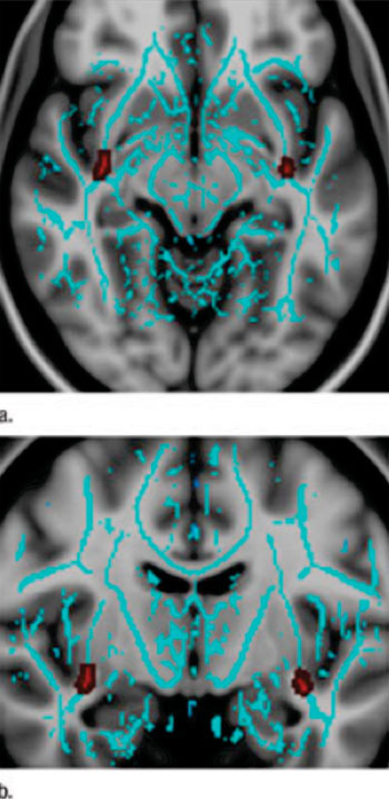
Image: Uncinate fasiculus, an important tract with the greatest concentration of progesterone receptors, show greater injury in males than females after mild traumatic brain injury (mTBI). (a) Axial and (b) coronal images show regions of decreased fractional anisotropy in male patients with mTBI relative to female mTBI patients, involving the uncinate fasiculus (red) bilaterally (Photo courtesy of Radiology).
Evaluating outcomes and recovery time after concussion can be very subjective. Typically, clinicians must rely on patient cooperation to assess injury severity. “MRI [magnetic resonance imaging] and CT [computed tomography] brain images of concussion patients are often normal,” said Saeed Fakhran, MD, assistant professor of neuroradiology at the University of Pittsburgh School of Medicine (PA, USA). “Diffusion tensor imaging is the first imaging technique that shows abnormalities associated with concussion, because it is able to see white matter tracts at a microscopic level.”
The investigators studied the medical records and imaging results of 69 patients diagnosed with mTBI between 2006 and 2013, including 47 males and 22 females, and 21 controls consisting of 10 males and 11 females (median age of males: 17; median age of females: 16). Of the 47 males with mTBI, 32 (68%) were injured while playing a sport, as were 10 of the 22 females (45%).
All patients underwent the same evaluation, including a computerized neurocognitive test and DTI of the brain. The DTI scans of the mTBI patients revealed abnormalities within the uncinate fasciculi (UF), a white matter tract that connects the frontal and temporal lobes of the brain. Although its precise role is controversial, the UF tract is believed to allow temporal lobe-based memory associations to modify behavior though interactions with another area of the brain.
The DTI scans revealed that compared to the female mTBI patients, the male mTBI patients had significantly decreased UF FA values. “In the future, we would like to look at the issue of gender and concussions more in depth to determine who does better and why,” Dr. Fakhran said.
A statistical analysis of the data revealed that UF FA value was a stronger predictor of recovery time than initial symptom severity based on neurocognitive testing. The most substantial risk factor for a recovery time longer than three months was decreased UF FA. Male gender also directly correlated with increased recovery time. “The potential of DTI and UF FA to predict outcome after concussion has great clinical impact,” Dr. Fakhran said. “Currently, we are heavily reliant on patient reporting, and patients may have ulterior motives, such as wanting to get back to play. But you can’t trick an MR scanner.”
The median time to symptom recovery for all concussion patients was 54 days. However, compared to the female patients who recovered in an average of 26.3 days, recovery substantially took longer for the male patients (an average of 66.9 days), regardless of the first symptom severity. “Male gender and UF FA values are independent risk factors for persistent postconcussion symptoms after three months and stronger predictors of time to recovery than initial symptom severity or neurocognitive test results,” Dr. Fakhran said.
Dr. Fakhran reported that the study’s findings indicate a potential role for UF FA values in triaging concussion patients in the future. “There’s prognostic value in DTI for both children participating in sports as well as for professional athletes,” he said. “Lower FA values in the uncinate fasciculi could offer a metric for evaluating the severity of mild traumatic brain injuries and predicting clinical outcome. We’re not at the point where DTI can provide individual prognoses yet, but that’s the hope and goal.”
Related Links:
University of Pittsburgh School of Medicine


.jpg)