Hyperpolarization MRI Scanning Technology Can Visualize Metabolic Changes
By MedImaging International staff writers
Posted on 06 Mar 2014
Danish researchers have revealed that a new scanning technique can visualize metabolic changes that have until now remained invisible, while they are actually occurring. Posted on 06 Mar 2014
The development of the hyperpolarization technique in 2003 by Danish scientists was a trailblazing moment that made it possible to see all the body’s cells with the help of a new contrast agent for magnetic resonance imaging (MRI) scans. The researchers have now taken another big leap towards a better determination of the body’s cells and with it also the developmental inner workings of the disease processes.
.")
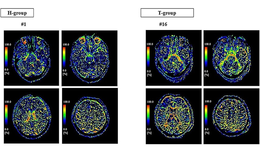
Image: Researchers can now follow metabolic changes by using a new MRI scanning technique. (Photo courtesy of Aurhus University).
“With the hyperpolarization method, sensitivity to specific contrast agents is up to 10,000 times higher than with a traditional MRI scanning. What we have now documented is that with the hyperpolarization MRI scanning we can not only see the differences in the metabolic patterns between healthy and ill, but we now can also see the metabolic changes as a consequence of acute influences while they are taking place in the diabetic kidneys. It is really groundbreaking,” said Dr. Christoffer Laustsen, an assistant professor from Aarhus University (Denmark).
This finding came from research of the connection between oxygen level and the development of kidney disease in rats with and without diabetes. The findings were published December 18, 2013, in the international journal Kidney International. Although the sophisticated hyperpolarization technology was first utilized to validate metabolic changes in the kidneys, it will be possible to use it to glean more clues into the development of diseases in all of the body’s organs.
Dr. Laustsen, noted, “With this method we will have a fingerprint of the cells and we will be able to follow whether these fingerprints change over time, regardless of which organs we examine. We will, for example, be able to see whether complications related to diabetes and cardiovascular diseases are emerging, or how a cancer tumor develops. And with greater knowledge of what is always going on at the cellular level we will be able to make a diagnosis earlier than we can today, and we will also be able to tailor treatment to the individual patient to a greater extent.”
The benefits of hyperpolarization scanning is not only that it provides in much more precise detail about what is occurring in the body. The new technology can also demonstrate itself to be very important specifically for the smallest patients. “Another great advantage of this method is that this is quite harmless. The contrast agent for the hyperpolarization scanning is sugar-based, so there is no risk of the body tissues being exposed to radiation. It will therefore be possible to scan children more often than today,” said Dr. Laustsen.
The studies of metabolic changes at the cellular level have so far been carried out on rats. However, Dr. Laustsen expects that researchers in Denmark will be ready to offer diagnostic examination of patients within two to three years.
Related Links:
Aarhus University

Digital Color Doppler Ultrasound System
MS22Plus

Computed Tomography System
Aquilion ONE / INSIGHT Edition

New
Digital Radiography System (Ceiling Free)
Digix CF Series

Ultrasonic Pocket Doppler
SD1