Sugar Lights Cancer Cells in MRI Scanners
By MedImaging International staff writers
Posted on 11 Jul 2013
A new study suggests that cancerous tumors can be detected by imaging the consumption of sugar with magnetic resonance imaging (MRI).Posted on 11 Jul 2013
Researchers at University College London (UCL; United Kingdom) developed a noninvasive technique called the glucose chemical exchange saturation transfer (glucoCEST) for MRI imaging of glucose uptake in vivo in a murine model, based on the exchange of protons between hydroxyl groups and water. The method differs from existing molecular imaging methods since it permits detection of the delivery and uptake of a metabolically active compound in physiological quantities.

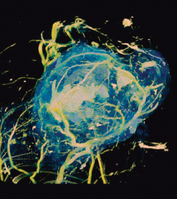
Image: MRI detecting the uptake of sugar in tumors (Photo courtesy of UCL).
The glucoCEST technique has been found to be sensitive to tumor glucose accumulation in colorectal tumor models, and can distinguish tumor types with differing metabolic characteristics and pathophysiologies. The method uses an injection of normal sugar and could offer a cheap, safe alternative to existing methods for detecting tumors, which require the injection of radioactive material. The study was published online ahead of print on July 7, 2013, in Nature Medicine.
“Our research reveals a useful and cost-effective method for imaging cancers using MRI – a standard imaging technology available in many large hospitals,” said senior author Prof. Mark Lythgoe, PhD, director of the UCL Center for Advanced Biomedical Imaging (CABI). “In the future, patients could potentially be scanned in local hospitals, rather than being referred to specialist medical centers.”
“Our cross-disciplinary research could allow vulnerable patient groups such as pregnant women and young children to be scanned more regularly, without the risks associated with a dose of radiation,” added senior author Prof. Xavier Golay, PhD.
Most cancer cells predominantly produce energy by a high rate of glycolysis followed by lactic acid fermentation in the cytosol, rather than by comparatively low rate of glycolysis followed by oxidation of pyruvate in mitochondria, as in most normal cells. Tumors, therefore, have a greater reliance on anaerobic glycolysis for energy production than normal tissues, resulting in glycolytic rates up to 200 times higher than those of their normal tissues of origin—a phenomenon that is known as the Warburg effect.
Related Links:
University College London


 Guided Devices.jpg)


