MRI Contrast Agents Not Associated with Parkinson’s Disease
By MedImaging International staff writers
Posted on 21 Jul 2016
A new study found no connection between gadolinium exposure and Parkinson’s disease (PD), a degenerative disorder characterized by tremor and impaired muscular coordination.Posted on 21 Jul 2016
Researchers at Researchers at Western University (WU; London, Canada) reviewed multiple linked administrative databases from Ontario (Canada) that included 246,557 patients older than 66 years who underwent at least one magnetic resonance imaging (MRI) scan between April 2003 and March 2013. Patients who were exposed to gadolinium-enhanced MRIs were compared with patients who received non-gadolinium-enhanced MRIs. MRI scans of the brain or spine were excluded.

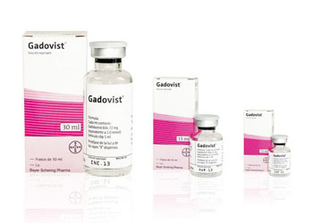
Image: Gadovist, a gadolinium-based contrast agent (Photo courtesy of Bayer).
The study found that 40.5% of the study patients received at least one dose of a gadolinium-based contrast agent (GBCA), with 81.5% undergoing a single study, and 2.5% receiving four or more GBCA-enhanced studies. In all, incident Parkinsonism developed in 1.2% of unexposed patients, and an equal 1.2% developed in those exposed to gadolinium. The study was published on July 5, 2016, in JAMA.
“Gadolinium-based contrast agents are used for enhancement during MRI; safety concerns have emerged over retained gadolinium in the globus pallidi,” concluded lead author Blayne Welk, MD, MSc, of Western University, and colleagues. “This result does not support the hypothesis that gadolinium deposits in the globus pallidi lead to neuronal damage manifesting as Parkinsonism. However, reports of other nonspecific symptoms) after gadolinium exposure require further study.”
Gadolinium--a rare earth heavy metal--is used for enhancement during MRI. Neurotoxic effects have been seen in animals, and when a GBCA is given intrathecally in humans. In July 2015, the U.S. Food and Drug Administration (FDA) stated that it was unknown whether gadolinium deposits in the globus pallidi, a subcortical structure of the brain, were harmful. The globus pallidus is a major component of the basal ganglia core along with the striatum and its direct target, the substantia nigra, and also retains close functional ties with the subthalamus, all part of the extrapyramidal motor system.
Related Links:
Western University


.jpg)

