Medical Imaging Breakthrough to Revolutionize Cancer and Arthritis Diagnosis
Posted on 03 Oct 2024
Photoacoustic tomography (PAT) imaging uses laser-generated ultrasound waves to detect subtle changes in small veins and arteries, typically less than a millimeter in size and up to 15mm deep in human tissues. However, traditional PAT technology has been too slow to produce high-quality 3D images suitable for clinical use. During a PAT scan, patients must remain completely still, and any movement during the slow scan can blur images, reducing their clinical value. Now, a new handheld scanner has been developed that can generate highly detailed 3D photoacoustic images in just seconds. This breakthrough opens the door for the use of PAT in clinical settings for the first time, potentially allowing earlier diagnosis of diseases. Conventional PAT scanners take more than five minutes to capture an image. By reducing the scanning time to a few seconds, the new technology improves image quality and makes the process much more practical for frail or unwell patients.
In the study, published in Nature Biomedical Engineering, the research team at University College London (London, UK) demonstrated that their new technology can deliver PAT imaging scans in real-time. This provides clinicians with accurate and detailed images of blood vessels, enhancing patient care. The team suggests that pending further testing, the scanner could help diagnose conditions such as cancer, cardiovascular disease, and arthritis within three to five years. The major breakthrough in this study is the dramatic reduction in imaging time, which is 100 to 1,000 times faster than previous PAT scanners. This speed prevents motion-induced blurring, enabling the production of highly detailed images of a quality unmatched by other scanners. The ability to acquire images in real time also makes it possible to observe dynamic physiological events. These advancements make the system suitable for clinical use, allowing doctors to examine aspects of human biology and disease that were previously inaccessible.
")
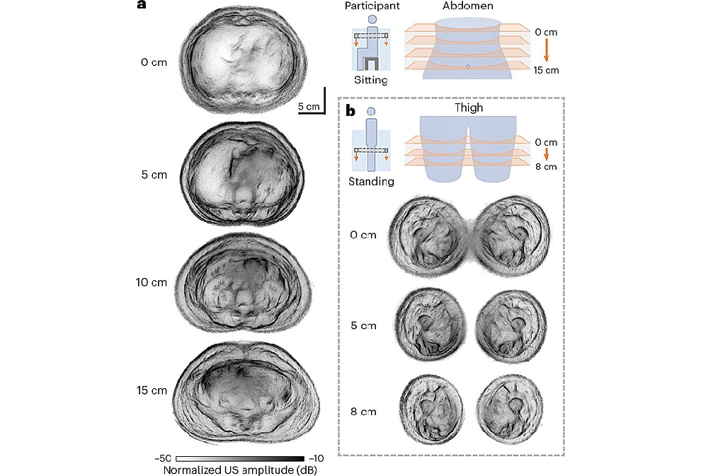
One key potential application for the new scanner is in the assessment of inflammatory arthritis, which requires scanning all 20 finger joints in both hands. The new scanner can perform this task in just a few minutes, compared to nearly an hour with older PAT scanners—an impractical time frame for elderly or frail patients. During pre-clinical testing, the team used the scanner on 10 patients with type-2 diabetes, rheumatoid arthritis, or breast cancer, as well as seven healthy volunteers. In three patients with type-2 diabetes, the scanner produced detailed 3D images of the microvasculature in their feet, revealing deformities and structural changes in the vessels. The scanner was also used to visualize skin inflammation related to breast cancer.
For conditions like peripheral vascular disease (PVD), a diabetes-related complication, early changes in tiny blood vessels are often invisible with conventional imaging techniques such as MRI. However, PAT can detect these early signs, offering the potential for earlier treatment to prevent tissue damage, poor wound healing, and the need for amputation. PVD affects more than 25 million people across the US and Europe. Similarly, in cancer cases, tumors often have a dense network of small blood vessels that are too small to detect with other imaging methods. PAT could be used to detect tumors and monitor their development more effectively. It may also assist surgeons in distinguishing tumor tissue from healthy tissue by visualizing the tumor's blood vessels, helping ensure complete removal during surgery and reducing the risk of recurrence. A key advantage of PAT technology is its sensitivity to hemoglobin, a light-absorbing molecule that produces ultrasound waves, making it especially effective in detecting blood vessels.
“One of the complications often suffered by people with diabetes is low blood flow in the extremities, such as the feet and lower legs, due to damage to the tiny blood vessels in these areas. But until now we haven’t been able to see exactly what is happening to cause this damage or characterize how it develops,” said Andrew Plumb, Associate Professor of Medical Imaging at UCL and consultant radiologist at UCLH and a senior author of the study. “In one of our patients, we could see smooth, uniform vessels in the left foot and deformed, squiggly vessels in the same region of the right foot, indicative of problems that may lead to tissue damage in future. Photoacoustic imaging could give us much more detailed information to facilitate early diagnosis, as well as better understand disease progression more generally.”