New PE-RADS Framework Standardizes Pulmonary Embolism Imaging Reports
Posted on 23 Jul 2026
Pulmonary embolism is a sudden blockage of the lung’s blood vessels that reduces pulmonary blood flow, can strain the right hear,t and may be fatal if untreated. Patients often present with shortness of breath, chest pain and low blood pressure, and rapid, consistent imaging assessment is essential. Variability in reporting across centers can impede coordination among emergency, radiology and critical care teams. To help address this challenge, a standardized imaging reporting framework for acute pulmonary embolism has been introduced.
The Pulmonary Embolism Reporting and Data Systems (PE-RADS) v2026 was developed by the American College of Radiology (ACR) in collaboration with representatives of six other medical groups. The framework was published in Radiology, a journal of the Radiological Society of North America (RSNA). It is designed to streamline communication and guide next steps in care for this potentially life‑threatening condition.
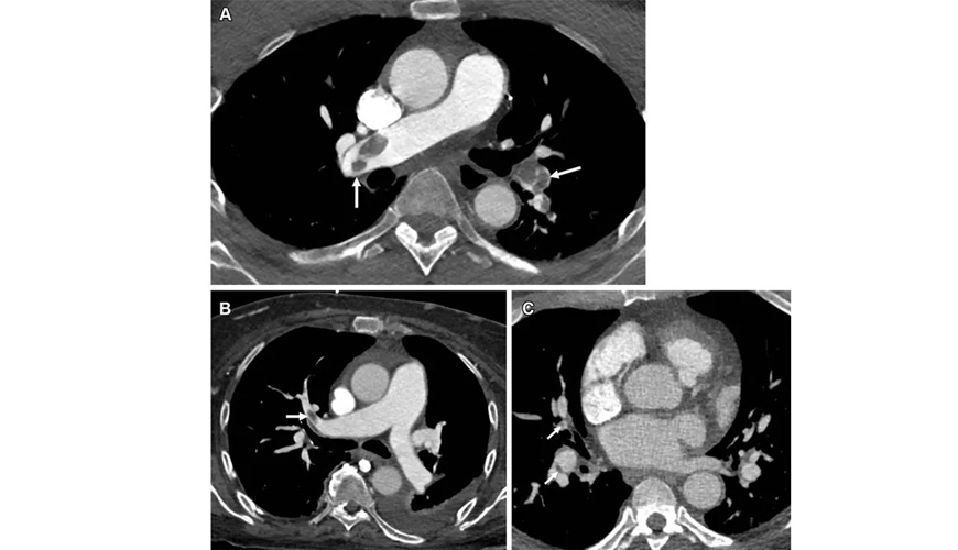
PE-RADS provides a hierarchical 0–4 grading scale to classify the most proximal clot location and incorporates right heart imaging features to support risk stratification and management planning. Modifiers indicate right ventricular (RV) enlargement and thrombus-in-transit when present, as well as limitations related to image quality and non–pulmonary embolism causes of pulmonary arterial disease. For example, “RV+” denotes RV enlargement, defined as an RV-to-left ventricle ratio of at least 1.0, while “T+” indicates definitive thrombus in the right atrium or RV.
CT pulmonary angiography is identified as the gold standard for diagnosing pulmonary embolism, while magnetic resonance angiography is described as a noninvasive, highly accurate alternative. The framework is aligned with existing pulmonary embolism guidelines to support adoption across multidisciplinary teams and is intended to evolve as evidence on outcomes and resource utilization emerges. PE-RADS was created within the ACR RADS program, which originated in 1992 to standardize disease-oriented imaging interpretation and reporting.
PE-RADS was created by radiologists and clinicians involved in evaluating and treating patients with suspected or confirmed pulmonary embolism. The ACR appointed half of the expert panel, while the remaining members were nominated by the American College of Emergency Physicians, American College of Chest Physicians, Pulmonary Embolism Response Team Consortium, Society of Thoracic Radiology, Society for Cardiovascular Angiography and Interventions, and Society of Interventional Radiology.
"The goal of PE-RADS is to improve communication between healthcare providers who care for patients with acute pulmonary embolism,” said Lynne M. Koweek, M.D., professor of radiology and associate professor of medicine at Duke University School of Medicine in Durham, North Carolina, and chair of the ACR PE-RADS committee. “As the first standardized lexicon and structured reporting system for pulmonary embolism imaging, PE-RADS v2026 is designed to provide a framework for reporting CT and MR angiographic findings in a clear, consistent manner to guide next steps in patient care.”
Related Links
RSNA