Simple 2D Ultrasound Tool Accurately Estimates Placental Volume
Posted on 16 Jul 2026
Stillbirth remains a major cause of perinatal loss, with about 21,000 cases annually in the United States. Although placental abnormalities are linked to adverse pregnancy outcomes, routine monitoring of placental size is not common in prenatal care. Accurate and practical measurement tools are needed to identify pregnancies with a disproportionately small placenta before complications arise. A new study shows that a simple two-dimensional ultrasound approach can provide accurate placental volume estimates using widely available equipment.
The Estimated Placental Volume (EPV) method offers a streamlined way to approximate placental size using standard two-dimensional ultrasound. Investigators at Yale School of Medicine assessed EPV against Virtual Organ Computer-aided Analysis (VOCAL), the three-dimensional ultrasound technique typically used to calculate placental volume. The peer-reviewed findings were published in the Journal of Perinatal Medicine in 2026.
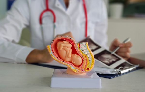
EPV relies on routine imaging and requires minimal operator training. During a scan, clinicians record placental width, height, and thickness and enter these values into a predefined equation to estimate total volume. The measurement process takes about 30 seconds, allowing it to be incorporated quickly into prenatal workflows without specialized equipment.
Investigators conducted a head-to-head comparison in 58 patients with healthy pregnancies and acquired two-dimensional and three-dimensional ultrasounds during the same clinical visit. Placental volume was calculated from the two-dimensional images using EPV and from the three-dimensional images using VOCAL. The resulting estimates showed a near-perfect correlation, indicating that EPV produced values almost identical to those generated by VOCAL. Researchers from Yale School of Medicine collaborated with the Pregnancy Research Branch of the Eunice Kennedy Shriver National Institute of Child Health and Human Development.
The authors noted that a practical and accurate gauge of placental size could encourage wider clinical evaluation of placental monitoring and enable earlier assessment during pregnancy. A follow-up analysis is being planned to identify which prenatal ultrasounds might have benefited from EPV measurement to detect small placentas. Advocacy efforts are also calling for routine placental measurement to be considered during standard prenatal care.
“Obstetricians have monitored fetal growth for decades, yet we have lacked a practical method to monitor the growth of the organ that supports fetal life. This study shows that the estimated placental volume obtained with 2D ultrasound provides placental size estimates comparable to those obtained with a more complex 3D method. We hope it will enable the studies needed to determine whether abnormalities in placental growth can identify pregnancies at risk before clinical complications develop,” said Roberto Romero, chief of the Pregnancy Research Branch in the Division of Intramural Research of the Eunice Kennedy Shriver National Institute of Child Health and Human Development.
Related Links
Yale School of Medicine