Cardiac MRI Detects Hidden Heart Dysfunction After Heart Attack
Posted on 10 Jul 2026
Myocardial infarction often leaves patients with persistent ventricular dysfunction that can progress without obvious signs. Detecting early decline remains difficult because standard measures may miss subtle abnormalities outside the scarred zone. Delayed recognition increases the risk of heart failure and rehospitalization. To help address this challenge, researchers have developed a cardiac MRI-based approach that interrogates mechanical changes in ventricular regions remote from the infarct.
Investigators from the INCLIVA Health Research Institute led a multicenter effort with the Clinical University Hospital of València, the University of Valencia, the Universitat Politècnica de València, the Cardiac Imaging Unit of Ascires Biomédico Group, and the Ascires–UPV Joint Research Unit. The team evaluated patients with chronic myocardial infarction using cardiac magnetic resonance imaging with strain analysis derived by feature tracking. The goal was to reveal dysfunction in apparently healthy myocardial segments that are distant from the index scar.
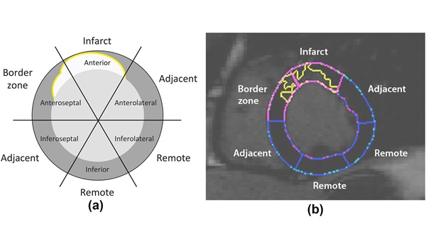
Feature tracking quantifies myocardial deformation through the cardiac cycle on routine cine images. By capturing subtle changes in strain, the technique can expose early functional impairment that is not evident on conventional parameters. In the study, basal ventricular segments were categorized into infarct, border zone, adjacent, and remote regions to localize strain abnormalities relative to scar distribution.
The researchers leveraged previously established normal reference values for magnetic resonance–derived strain from a healthy population. Using these benchmarks, they compared patient results to identify small deviations in remote myocardium that may precede global deterioration. The approach seeks to improve prognostic assessment and enable earlier, more personalized therapy before symptoms or overt remodeling occur.
The study was published in Clinical Radiology. According to the team, identifying remote ventricular dysfunction could refine risk stratification after myocardial infarction and support precision follow-up in cardiology clinics.
“Having these normal values available made it possible, in the present study, to accurately compare the results obtained in patients with chronic infarction and detect subtle deviations that would not be apparent using conventional techniques. This approach allows for a more precise and quantitative assessment of cardiac function, overcoming the limitations of traditional measures such as ejection fraction,” said David Moratal, professor in the Department of Electronic Engineering at the Universitat Politècnica de València.