AI Tool Predicts Side Effects from Lung Cancer Treatment
Posted on 03 Mar 2026
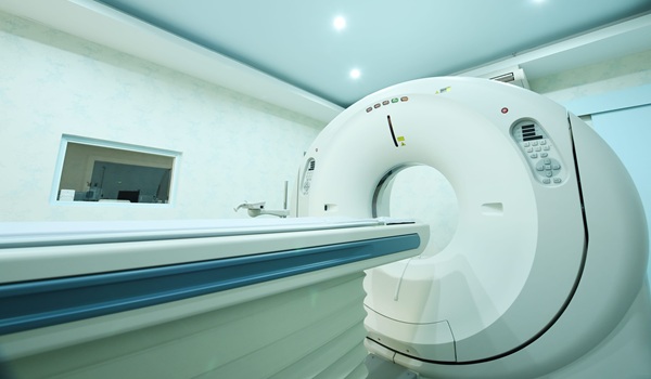
Radiation therapy is a central treatment for lung cancer, but even carefully targeted radiation can affect surrounding healthy tissue. Patients may develop side effects such as lung inflammation, coughing, and shortness of breath when nearby lung structures receive unintended exposure. Predicting which patients are most vulnerable to radiation-related lung toxicity remains a challenge before treatment begins. Researchers have now developed a machine learning tool that identifies lung lobes on CT scans to help estimate side-effect risk more accurately.
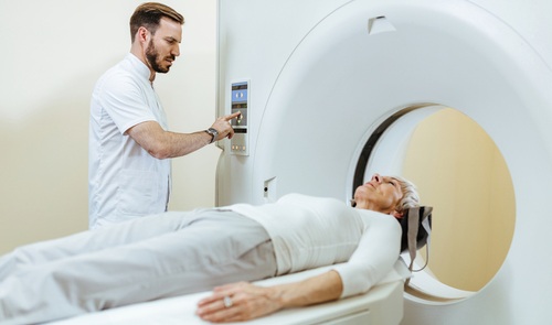
In a new study, researchers at Sidney Kimmel Medical College (Philadelphia, PA, USA) focused on analyzing the lung’s five natural subunits, or lobes, rather than treating the lung as a single organ during radiation planning. Traditionally, clinicians must manually outline each lobe on CT scans to determine radiation dose distribution, a process that takes three to four hours per patient. To streamline this, the team trained a machine learning model using CT scans from 40 lung cancer patients across two institutions where lobes had already been carefully delineated by specialists.
")
The trained model was then tested on 10 previously unseen CT scans. The results showed that the artificial intelligence (AI) model could identify and outline all five lung lobes with accuracy comparable to that of clinicians. The study, published in Reports of Practical Oncology and Radiotherapy, demonstrated that the automated system performs the task in less than a second, compared to several hours required for manual contouring. This efficiency makes lobe-level dose analysis practical in routine clinical workflows.
By enabling rapid lobe-specific radiation assessment, the tool may improve the prediction of lung toxicity, particularly when the lower lobes receive higher radiation doses. Identifying high-risk patients before therapy begins could allow clinicians to adjust treatment plans and reduce the likelihood of serious complications. The approach highlights the growing role of AI in personalizing radiation oncology and optimizing patient safety. Future studies will further evaluate how incorporating lobe-level analysis influences treatment planning and outcomes.
“A machine learning model can identify lung lobes just as accurately as a clinician. But instead of hours, it takes less than a second,” said medical physicist Yevgeniy Vinogradskiy, PhD, who led the research team. “If we can identify which patients are at higher risk, we can intervene earlier and potentially prevent serious complications.”
Related Links:
Sidney Kimmel Medical College