New PET/CT Technique Accurately Detects Neuroblastoma in Children with Short Scan Time and No Anesthesia
Posted on 22 Aug 2024
Neuroblastoma, the most common extracranial solid tumor in children, has an overall survival rate of 70%. Traditionally, the 123I-MIBG SPECT/CT scanning procedure has been the standard of care for staging, monitoring response, and follow-up in these cases. This method involves a two-day protocol where sedation or general anesthesia is often necessary, especially as the procedure lasts over two hours and is primarily used for infants. Minimizing radiation exposure and avoiding sedation is critical for young patients. Researchers have now introduced a new molecular imaging method that utilizes a novel tracer combined with an advanced PET/CT scanner to identify neuroblastoma in children with high sensitivity, requiring a scan time of just minutes and no sedation or anesthesia.
Developed by specialists at Copenhagen University Hospital-Rigshospitalet (Copenhagen, Denmark), the 18F-MFBG LAFOV PET/CT method enables precise accurate diagnosis and aids in therapeutic decision-making for neuroblastoma in children. This technique employs the tracer 18F-MFBG and only requires a single-day protocol using a long-axial-field-of-view (LAFOV) PET/CT scanner, which is about 10 times more sensitive than conventional digital PET/CT scanners. Researchers conducted a comparative study using both the 123I-MIBG SPECT/CT and the new 18F-MFBG LAFOV PET/CT on 10 children with neuroblastoma, assessing the diagnostic accuracy and practicality of both methods.
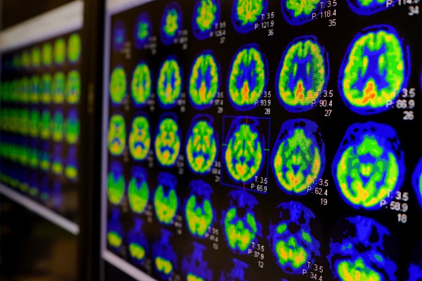
![Image: [18F]MFBG LAFOV PET/ULD CT (top) and [123I]MIBG scintigraphy with SPECT/LD CT images (bottom) of 7-wk-old girl with neuroblastoma (Photo courtesy of The Journal of Nuclear Medicine)](https://globetechcdn.com/mobile_medicalimaging/images/stories/articles/article_images/2024-08-22/JNM Aug 2024 - Borgwardt Figure 3A.jpg "Image: [18F]MFBG LAFOV PET/ULD CT (top) and [123I]MIBG scintigraphy with SPECT/LD CT images (bottom) of 7-wk-old girl with neuroblastoma (Photo courtesy of The Journal of Nuclear Medicine)")
The findings published in the August issue of The Journal of Nuclear Medicine showed that 18F-MFBG LAFOV PET/CT eliminated the need for sedation or anesthesia, with 80% of children requiring anesthesia under the traditional method. The new PET/CT procedure required only two minutes to acquire images free from motion artifacts, sufficient for producing clinically useful images. The 18F-MFBG LAFOV PET/CT also detected more lesions than the 123I-MIBG SPECT/CT in 80% of cases, with the remaining 20% showing equal lesion detection. Furthermore, the SIOPEN and Curie scores, which assess metastatic disease burden, indicated more extensive disease with the new PET/CT method. This technique also provided a more precise diagnosis of intraspinal, retroperitoneal lymph node, and bone marrow involvement.
“A scan with a much higher sensitivity can find very small lesions and the exact extension in the body and can be extremely beneficial in determining the right course of treatment,” said Lise Borgwardt, MD, PhD, senior consultant in pediatric nuclear medicine at Copenhagen University Hospital-Rigshospitalet. “The fact that these scans can be performed without anesthesia or sedation, and at a lower radiation dose is a big step forward for the children, parents, and the healthcare system in general.”
Related Links:
Copenhagen University Hospital-Rigshospitalet