AI-Enhanced Technology Predicts Fatal and Non-Fatal Cardiac Events from Routine CCTA
Posted on 20 Nov 2023
Annually, over 8 million people in the United States seek emergency medical attention due to chest pain. Typically, these patients undergo coronary computed tomography angiography (CCTA) to check for coronary artery disease (CAD), which is caused by the narrowing or obstruction of the arteries supplying blood to the heart. However, significant narrowing is not always detected during CCTA in many of these cases. Despite this, a considerable number of these individuals suffer fatal heart attacks in subsequent years due to minor, yet inflamed plaques that burst. Now, an innovative artificial intelligence (AI) technology has been developed that can predict both fatal and non-fatal cardiac events. This prediction is independent of standard clinical risk assessments and CCTA results, and it has the potential to revolutionize the way CAD is treated by transforming the risk evaluation and management of patients who undergo routine CCTA.
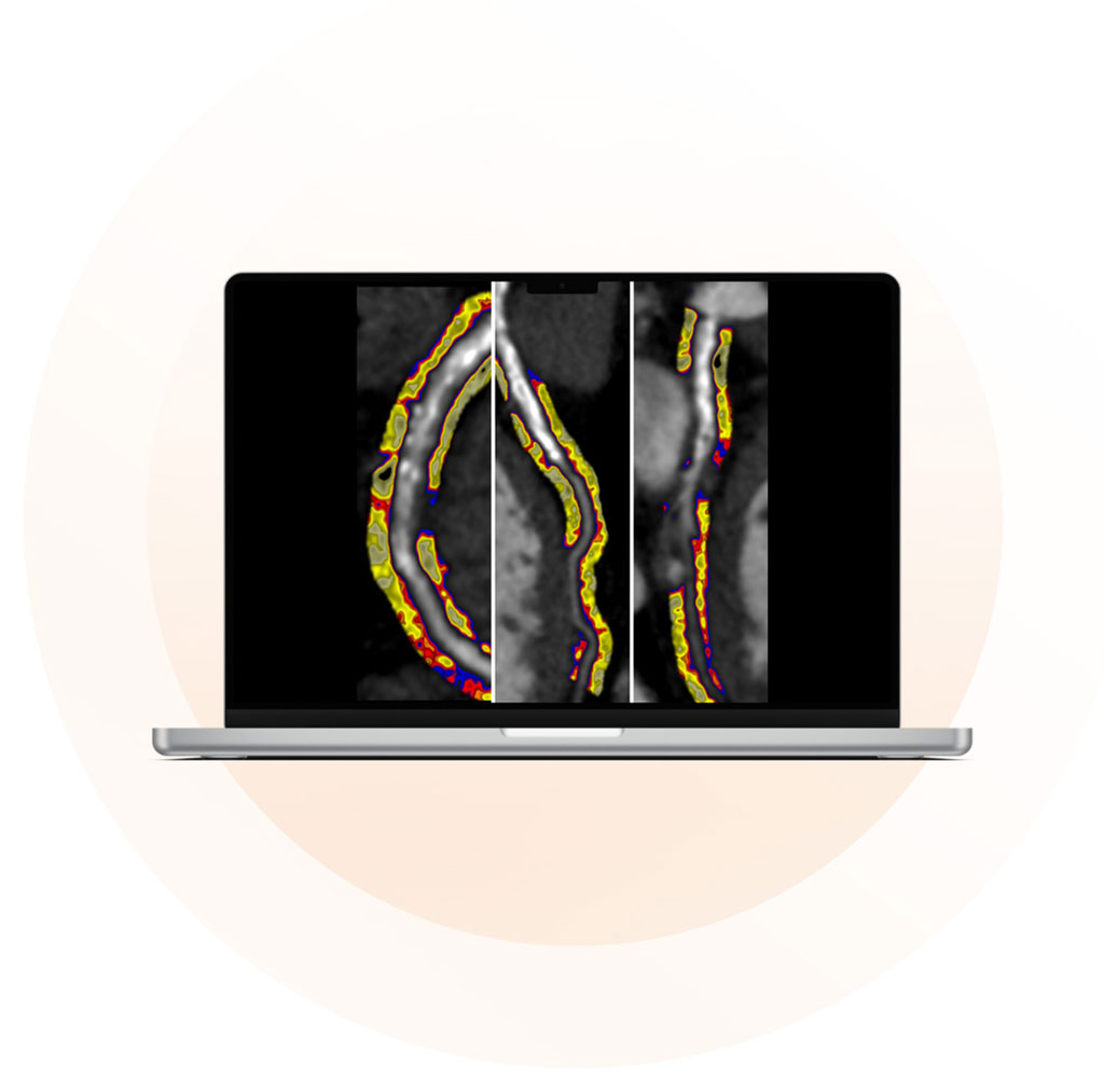
The CaRi-Heart technology from Caristo Diagnostics (Oxford, UK) is an AI-powered imaging tool that assesses coronary inflammation and characterizes plaque, aiding clinicians in better decision-making for patient care. This cloud-based software as a service (SaaS) medical device utilizes AI to process standard CCTA images. It introduces a unique imaging biomarker, the Fat Attenuation Index (FAI), to measure the extent of coronary inflammation and provides detailed analysis of plaque characteristics that are often linked with heart attacks.

The ORFAN study, the largest of its kind aimed at evaluating the effectiveness of CT imaging biomarkers in predicting long-term cardiovascular outcomes, demonstrated that coronary inflammation measured by Caristo's CaRi-Heart FAI-Score could foresee fatal and non-fatal cardiac events. These predictions were made independently of routine risk factors and standard CCTA interpretations. In this study, CaRi-Heart Risk functioned as the "AI-Risk" model, which reclassified about 30% of patients into higher risk categories and around 10% into lower risk categories. When these results were reviewed by clinicians, it led to changes in patient management in nearly half of the cases, primarily targeting previously unrecognized coronary inflammation.
"We are excited to see these exceptional clinical results enabled by our CaRi-Heart technology," said Frank Cheng, CEO of Caristo Diagnostics. "To eradicate heart disease as the number one cause of death globally, it is important for us to realize that people may have heart attack soon after a 'normal' CCTA test showing zero calcium score, no plaque, and no stenosis. The CaRi-Heart technology has the potential to save millions of lives worldwide by improving risk stratification based on CCTA-based coronary inflammation measurement. We look forward to introducing our technology across geographies to transform cardiac care and make heart attacks a preventable reality worldwide."
Related Links:
Caristo Diagnostics


