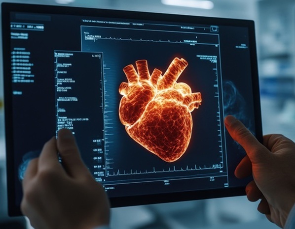
Ultrasound Can Image Immune Cells Enhanced With Microbubbles to Diagnose Early Stage Cancer
Posted on 02 Jun 2023
Macrophages, a type of white blood cell, protect the human body by surrounding and consuming foreign particles such as bacteria, viruses, and dead cells. Notably, these immune cells tend to gather within solid tumors, suggesting that monitoring their movement could yield innovative methods for early cancer and metastasis detection. Several imaging techniques including optical imaging, MRI, and PET scans have been employed to observe macrophages. However, each of these methods has its own shortcomings.
Optical imaging offers limited penetration depth, making it effective only for immune cells near the skin's surface. While MRI can provide detailed information about the macrophages and tumor surroundings, it has difficulty in detecting a small cell count. PET scans, despite their sensitivity, have a low resolution and require ionizing radiation. Now, a recent research breakthrough has demonstrated that attaching microbubbles to macrophages can generate detailed and sensitive tracking images to aid disease diagnosis.
")
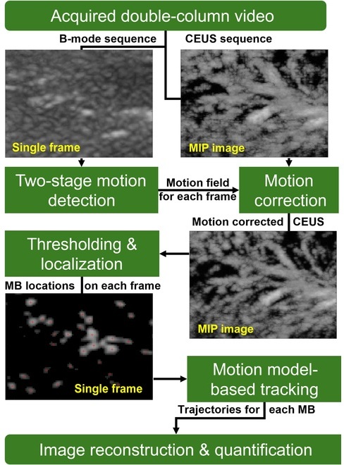
A research team at Georgia Institute of Technology (Atlanta, GA, USA) pioneered this approach by attaching microbubbles to macrophages, which allowed these cells to send back an echo when hit with ultrasound. This technique facilitated the high-resolution and sensitive visualization of macrophages in a living organism. In vivo macrophage visualization could also serve as a potent tool in understanding immune responses and monitoring the effectiveness of therapeutic treatments.
“Ultrasound could potentially overcome these limitations, as it is nonionizing, noninvasive, and has great depth of penetration,” said Ashley Alva of the Georgia Institute of Technology. “In the future, we envision harvesting macrophages from patients, labeling them with microbubbles, and reinserting them to improve diagnosis using ultrasound tracking methods and algorithms we are currently developing. If needed, the harvested macrophages could also be engineered to become more sensitive to certain disease characteristics before being reinjected into patients.”
Related Links:
Georgia Institute of Technology


.jpeg)