PET Scans Show Dynamic Function of Multiple Organs
By MedImaging International staff writers
Posted on 30 Jan 2020
A new study demonstrates a positron emission tomography (PET) image reconstruction method that helps researchers capture real-time videos of blood flow and heart function.Posted on 30 Jan 2020
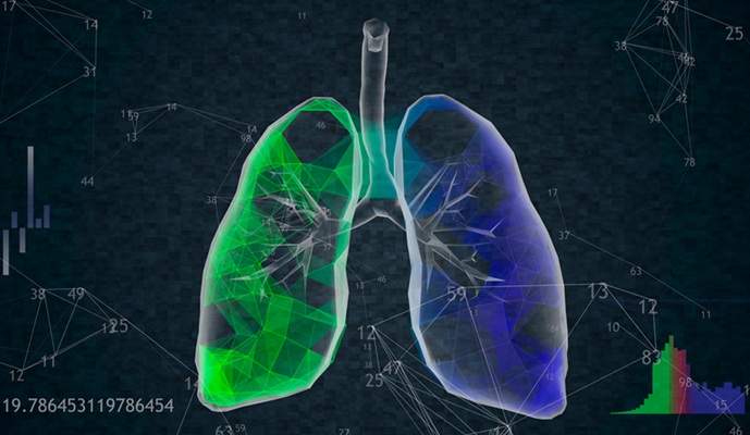
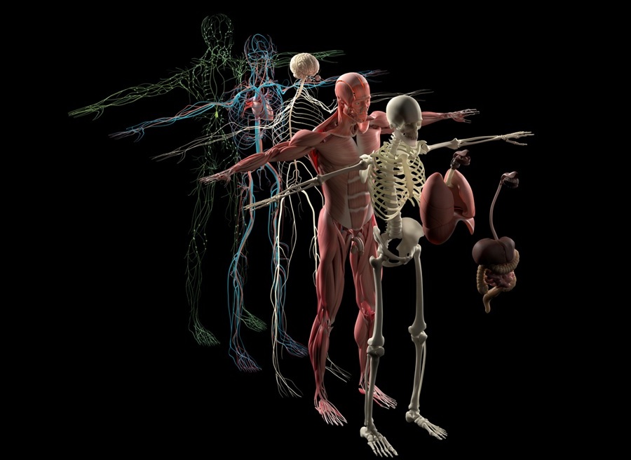
Researchers at the University of California Davis (UCD; USA) and Fudan University (Shanghai, China) have developed new methods to reduce noise and reconstruct video images from raw data of volunteers scanned by the Explorer, a full body PET and x-ray computed tomography (CT) scanner that can evaluate all body organs and tissues simultaneously. They were able to see changes on a scale of 100 milliseconds (one-tenth of a second), and use these to create high quality real-time movies of the scans.
")
Image: Full body PET/CT on the Explorer scanner (Photo courtesy of UCD)
For example, Explorer quantitatively measured blood flow, glucose uptake all over the body at the same time, cancer metastasis beyond the single tumor site, inflammation and infection, and immunological or metabolic disorders, as well as many other diseases. In one scan shown, a volunteer injected in with a short-lived radioactive tracer was scanned in real-time, showing the tracer moving up the body to the heart, flowing through the right ventricle to the lungs, back through the left ventricle and on to the rest of the body. Another video shows heart motion and cardiac contraction with clear delineation of the end-systolic and end-diastolic phases. The study was published on January 20, 2020, in Proceedings of the National Academy of Sciences (PNAS).
“It's a combination of the scanner and advanced data reconstruction methods that make this possible. The tradeoff between image quality, acquisition time, and injected radiation dose will vary for different applications,” said lead author Xuezhu Zhang , PhD, of UCD. “This has applications in real-time tracking of blood flow over the human circulatory system, motion-frozen heart beating, and breathing monitoring for cardiovascular and cerebrovascular disease and analysis of respiratory system function.”
PET is a nuclear medicine imaging technique that produces a 3D image of functional processes in the body. The system detects pairs of gamma rays emitted indirectly by a positron-emitting radionuclide tracer. Tracer concentrations within the body are then constructed into a 3D image by computer analysis. In modern PET-CT scanners, 3D imaging is often accomplished with the aid of a CT X-ray scan performed on the patient during the same session, in the same machine.
Related Links:
University of California Davis
Fudan University

Radiation Safety Barrier
RayShield Intensi-Barrier

Mammo DR Retrofit Solution
DR Retrofit Mammography

Ultrasonic Pocket Doppler
SD1

MRI System
nanoScan MRI 3T/7T