Stitching Software Creates Virtual 3D Tissue Maps
By MedImaging International staff writers
Posted on 21 Aug 2019
A new study shows how pioneering software reconstructs tissue slides into a navigable, three-dimensional (3D) organ model that can illustrate cellular functions such as gene expression.Posted on 21 Aug 2019
Developed by researchers at Ludwig-Maximilians-Universität (LMU; Munich, Germany), Max Delbrück Center for Molecular Medicine (MDC; Berlin, Germany), Howard Hughes Medical Institute (HHMI; Ashburn, USA), and other institutions, BigStitcher allows simple and efficient alignment of image datasets acquired by lightsheet, widefield, or confocal microscopes. Images of almost arbitrary size are supported, ranging from very small images up to volumes in the range of many terabytes.
.")
Image: A virtual cross-section of the complete nervous system of a fruit fly larva (Photo courtesy of Janelia/ MDC).
Dubbed by the researchers as “Google Maps in 3D”, BigStitcher enables interactive visualization, fast and precise alignment, spatially resolved quality estimation, real-time fusion, and deconvolution of dual-illumination, multi-tile, multiview datasets at any level of detail desired. The converted images can also be rotated and turned virtually, providing an overview of the entire sample, or zoomed into the level of individual structures, and even systematically characterized at the single-cell level. The software also compensates for optical effects, improving accuracy and enabling subsequent biological analysis.
Other features include integrated downstream processing of the image data; pre-selection of the best illumination direction at every image block in the sample; support of non-regular acquisition grids, which includes 'intelligent acquisitions' where some of the image blocks can be missing if only background is present; and support of image data acquired at different resolutions to combine overview scans with high resolution acquisitions of specific areas of interest. BigStitcher is being distributed within the Fiji framework, and can be downloaded and used free of charge. The study was published on August 5, 2019, in Nature Methods.
“One can not only get an overview of the big picture, but can also zoom in to specifically examine individual structures at the desired resolution,” said senior author Stephan Preibisch, PhD, head of the MDC research group on Microscopy, Image Analysis & Modeling of Developing Organisms. “The software automatically assesses the quality of the acquired data. The brighter a particular region of, say, a mouse brain or a human organ is displayed on screen, the higher the validity and reliability of the acquired data.”
Related Links:
Ludwig-Maximilians-Universität
Max Delbrück Center for Molecular Medicine
Howard Hughes Medical Institute

Gold Member
Solid State Kv/Dose Multi-Sensor
AGMS-DM+

New
Wireless Handheld Ultrasound System
TE Air
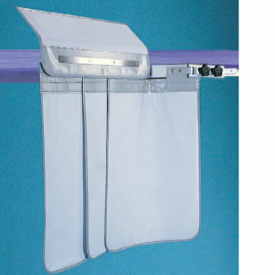
Under Table Shield
3 Section Double Pivot Under Table Shield
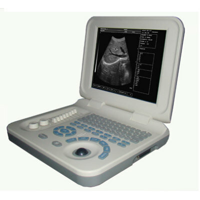
Laptop Ultrasound Scanner
PL-3018

.jpg)