New Artificial Intelligence Algorithm Boosts Quality of Brain Magnetic Resonance Imaging
By MedImaging International staff writers
Posted on 19 Feb 2020
Researchers from the ICAI Group – Computational Intelligence and Image Analysis – of the University of Malaga (Málaga, Spain) have designed a method that is capable of improving brain images obtained through magnetic resonance imaging using artificial intelligence (AI). The new model manages to increase image quality from low resolution to high resolution without distorting the patients' brain structures, using a deep learning artificial neural network – a model based on the functioning of the human brain – that "learns" this process.Posted on 19 Feb 2020
The algorithm developed by the UMA yields more accurate results in less time, with clear benefits for patients. The technique allows the activity of identification to be performed alone, without supervision; an identification effort that the human eye would be incapable of doing. According to the researchers, the results will enable specialists to identify brain-related pathologies, such as physical injuries, cancer or language disorders, among others, with increased accuracy and definition, as the image details are thinner, thus eliminating the need for performing additional tests when diagnoses are uncertain.
")
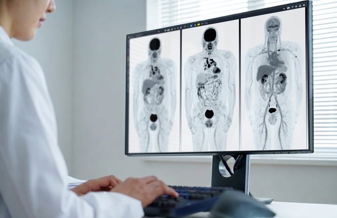
Image: Artificial intelligence to improve resolution of brain magnetic resonance imaging (Photo courtesy of University of Malaga)
"Deep learning is based on very large neural networks, and so is its capacity to learn, reaching the complexity and abstraction of a brain," said researcher Karl Thurnhofer, main author of the study. "So far, the acquisition of quality brain images has depended on the time the patient remained immobilized in the scanner; with our method, image processing is carried out later on the computer."
Related Links:
University of Malaga

Diagnostic Ultrasound System
DC-80A
 Guided Devices.jpg)
Ultrasound-Guided Biopsy & Visualization Tools
Endoscopic Ultrasound (EUS) Guided Devices

Mammo DR Retrofit Solution
DR Retrofit Mammography

Post-Processing Imaging System
DynaCAD Prostate