New AI Method Predicts Breast Cancer Five Years in Advance
By MedImaging International staff writers
Posted on 18 May 2019
Researchers from two major institutions have developed a new tool with advanced artificial intelligence (AI) methods to predict a woman’s future risk of breast cancer. The currently available models that use factors such as family history and genetics fall far short in predicting an individual woman’s likelihood of being diagnosed with breast cancer.Posted on 18 May 2019
In some models, breast density—the amount of dense tissue compared to the amount of fatty tissue in the breast on a mammogram— has been added to improve risk assessment as it is an independent risk factor for breast cancer. Since it is based on subjective assessment that can vary across radiologists, deep learning, a subset of AI in which computers learn by example, has been studied as a way to standardize and automate these measurements.
.")
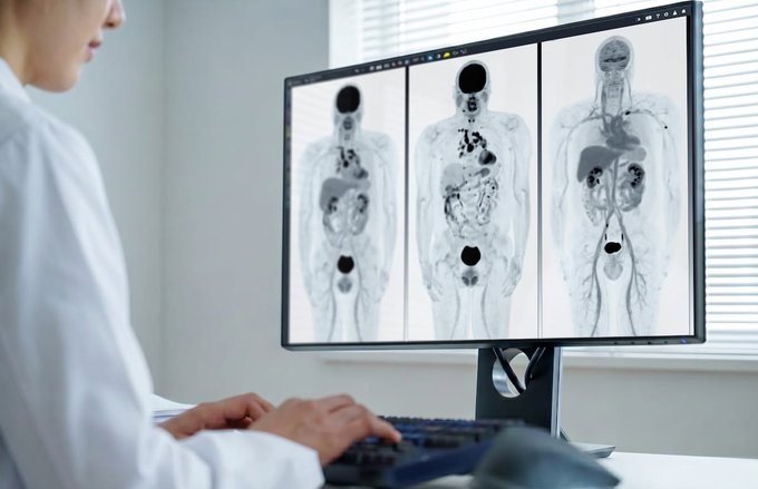
Image: A new AI method for detecting breast cancer is expected to surpass existing methods that fall short in their predictions (Photo courtesy of MIT).
Adam Yala, a Ph.D. candidate at the Massachusetts Institute of Technology (MIT), in collaboration with Regina Barzilay, Ph.D., an AI expert and professor at MIT, and Constance Lehman, M.D, Ph.D., chief of breast imaging at Massachusetts General Hospital and professor of radiology at Harvard Medical School, recently compared three different risk assessment approaches.
The first model relied on traditional risk factors, the second on deep learning that used the mammogram alone, and the third on a hybrid approach that incorporated both the mammogram and traditional risk factors into the deep learning model. The researchers used nearly 90,000 full-resolution screening mammograms from about 40,000 women to train, validate and test the deep learning model. They were able to obtain cancer outcomes through linkage to a regional tumor registry.
The deep learning models yielded substantially improved risk discrimination over the Tyrer-Cuzick model, a current clinical standard that uses breast density in factoring risk. When comparing the hybrid deep learning model against breast density, the researchers found that patients with non-dense breasts and model-assessed high risk had 3.9 times the cancer incidence of patients with dense breasts and model-assessed low risk. The advantages held across different subgroups of women.
“There’s much more information in a mammogram than just the four categories of breast density. By using the deep learning model, we learn subtle cues that are indicative of future cancer,” said Yala. “There’s a very large amount of information in a full-resolution mammogram that breast cancer risk models have not been able to use until recently. Using deep learning, we can learn to leverage that information directly from the data and create models that are significantly more accurate across diverse populations.”
“Unlike traditional models, our deep learning model performs equally well across diverse races, ages and family histories,” Dr. Barzilay said. “Until now, African-American women were at a distinct disadvantage in having accurate risk assessment of future breast cancer. Our AI model has changed that.”
“A missing element to support more effective, more personalized screening programs has been risk-assessment tools that are easy to implement and that work across the full diversity of women whom we serve,” Dr. Lehman said. “We are thrilled with our results and eager to work closely with our health care systems, our providers and, most importantly, our patients to incorporate this discovery into improved outcomes for all women.”
Related Links:
MIT

Silver Member
X-Ray QA Device
Accu-Gold+ Touch Pro

Digital Intelligent Ferromagnetic Detector
Digital Ferromagnetic Detector

Digital Radiography System
DR-300

Digital Radiographic System
OMNERA 300M