AI Software Diagnoses Lung Cancer from CT Images
By MedImaging International staff writers
Posted on 07 Mar 2019
A team of Russian researchers, in collaboration with radiologists, has developed an intelligent software system for lung cancer diagnostics. The software, which can be installed on any computer, analyzes patients' computed tomography (CT) results within 20 seconds and provides an image in which the pathology is clearly marked. The system has been named Doctor AIzimov (AI for Artificial Intelligence) in honor of the science-fiction writer Isaac Asimov, who developed three famous laws of robotics.Posted on 07 Mar 2019
The first tests of this intelligent system were carried out at the end of 2018 in which the system analyzed CT images of 60 patients and found focal nodules in lungs of small sizes (2 mm). The open testing of the intelligent system will be carried out at the beginning of 2019. The system will be adapted to analyze the results of the ultrasound and X-ray medical investigation of other organs.
.")
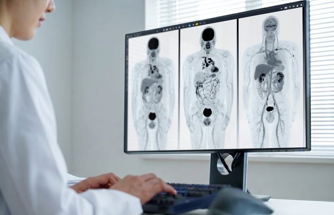
Image: The Doctor AIzimov system analyzes computed tomography results within 20 seconds (Photo courtesy of Peter the Great Saint-Petersburg Polytechnic University).
In a new proposed and developed approach to the lung cancer classification using the chord method, segmented CT images are used: points are randomly drawn on the surface of a nodule, after that the points are connected by lines (chords). The length histogram of the chords reflects the shape and structure of the tumor. Although the system examines every nodule from the inside, its external surroundings are also very important. To learn more about the tumor, it is placed in a cube, and perpendiculars are drawn from its edges to the surface of the nodule.
Thus, instead of classifying graphically complex and heavy images of the CT (the size of every CT image is approximately 1 GB), the nodule is represented in the form of compact and simple histograms, which are then analyzed by the Doctor AIzimov system. The scientists have also trained the system to distinguish malignant and benign tumors. Currently, the dataset holds CT images of about 250 patients and the scientists plan to increase the number of images by four times by mid-2019.
With each new CT image, the system self-improves. In the future, a patient's CT images will be transferred to the supercomputer using the Internet that will reduce the diagnostic testing time per patient from 20 till 2 seconds. After that a radiologist will receive the marked image instead of the large CT image, thus significantly reducing the time needed for the analysis and diagnostics.
“Many different objects may be detected on the CT images, so the main task was to train the system to recognize what each of the objects represents. Using the clinical and radiological classification, we are trying to train the system not only to detect tumors, but also to distinguish other diseases similar to cancer,” said Anna Meldo, the head of the Radiology Department of the St. Petersburg Clinical Research Center for Specialized Types of Medical Care (Oncological).

Computed Tomography System
Aquilion ONE / INSIGHT Edition

Mobile X-Ray System
K4W

Digital Radiography System
DR-300

Medical Radiographic X-Ray Machine
TR30N HF