ECG Imaging Algorithm Could Help Reduce Invasive Heart Procedures
By MedImaging International staff writers
Posted on 30 Oct 2018
A group of researchers from the University of California, Santa Barbara (Santa Barbara, CA, USA) have developed new algorithms to localize the source of an atrial fibrillation, an abnormal heart rhythm.Posted on 30 Oct 2018
Doctors use invasive procedures to map the hearts of patients suffering from atrial fibrillation and decide whether an ablation procedure to remove heart tissue is likely to have a positive outcome. Computed tomography (CT) scans or ultrasounds are useful in determining the structure of a patient’s heart, although invasive electrical procedures are used to identify and localize the source of the atrial fibrillation.

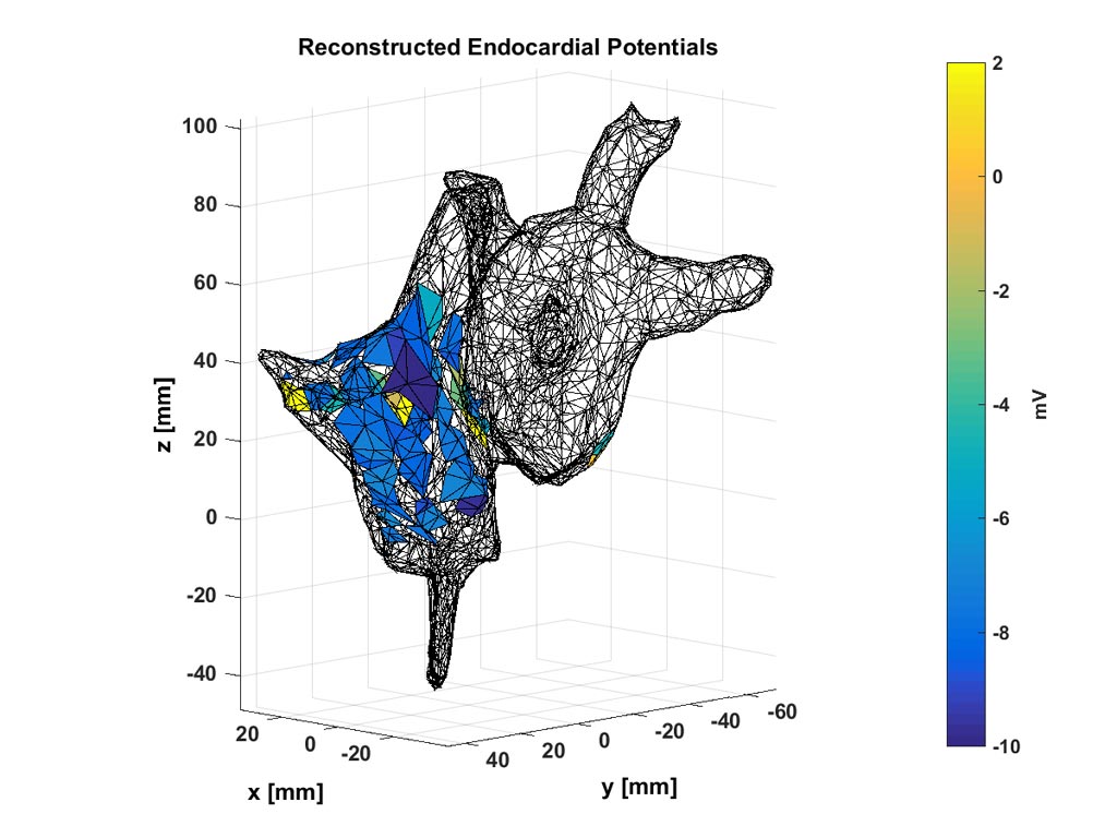
Image: Doctors can use these noninvasive maps of electrochemical potentials inside a patient\'s heart to localize the source of an abnormal heart rhythm (Photo courtesy of Abhejit Rajagopal).
The new algorithms are based on the concept that the inverse operator, a function that maps body-surface electrocardiogram signals to endocardial potentials, can be non-linear and optimized using a set of historical data. This allows them to learn a model for predicting cardiac potentials from electrocardiograms that are realistic, accurate, and amenable to general-purpose use as a new cardiac imaging tool. This is significant because it suggests that much higher resolution reconstruction is possible if non-linear reconstruction algorithms are used, as compared to what is theoretically known using linear methods and partial data.
“Imagine a world where instead of a doctor listening to your heart through a stethoscope they can see a live video of your heart beating via ultrasound with corresponding electrical measurements of the local potentials on or around the cardiac tissue,” said UC Santa Barbara graduate student Abhejit Rajagopal, author of the paper published in the journal APL Bioengineering, from AIP Publishing. “The goal is for doctors to be able to treat patients with cardiac issues without needing to use invasive surgeries just to determine the cause.”
Related Links:
University of California, Santa Barbara


