New AI Technique Dramatically Improves Quality of Medical Imaging
By MedImaging International staff writers
Posted on 05 Apr 2018
Researchers have developed a new technique based on artificial intelligence (AI) and machine learning that enables radiologists to acquire higher quality images without having to collect additional data at the cost of increased radiation dose for computed tomography (CT) and positron emission tomography (PET) or uncomfortably long scan times for magnetic resonance imaging (MRI).Posted on 05 Apr 2018
The technique named AUTOMAP (automated transform by manifold approximation) marks a significant step forward for biomedical imaging. Researchers from the Athinoula A. Martinos Center for Biomedical Imaging at Massachusetts General Hospital (MGH) developed the technique by taking advantage of the various strides made in recent years in the neural network models used for AI and in the graphical processing units (GPUs) that drive the operations. This is because image reconstruction, particularly in the context of AUTOMAP, requires an immense amount of computation, particularly during the training of the algorithms. The availability of large datasets ("big data") required to train large neural network models such as AUTOMAP was another important factor that helped researchers to develop this technique.
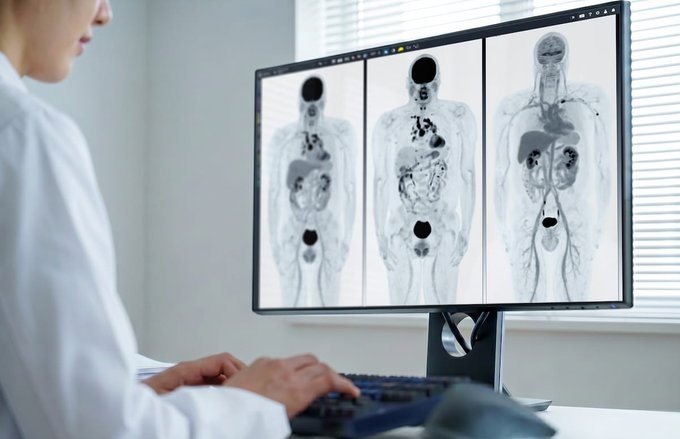
 and AUTOMAP (right) (Photo courtesy of Athinoula A. Martinos Center for Biomedical Imaging, Massachusetts General Hospital).")
Image: A new artificial-intelligence-based approach to image reconstruction – called AUTOMAP – yields higher quality images from less data, reducing radiation doses for CT and PET and shortening scan times for MRI. Shown here are MR images reconstructed from the same data with conventional approaches (left) and AUTOMAP (right) (Photo courtesy of Athinoula A. Martinos Center for Biomedical Imaging, Massachusetts General Hospital).
In addition to producing high-quality images in less time with MRI or with lower doses with X-ray, CT and PET, AUTOMAP offers several potential benefits for clinical care. For instance, its processing speed can help the technique aid in real-time decision making about imaging protocols while the patient is in the scanner. The technique can also help in advancing other AI and machine learning applications. Since most of the current excitement surrounding machine learning in clinical imaging is focused on computer-aided diagnostics, AUTOMAP could play a role in advancing them for future clinical use as these systems rely on high-quality images for accurate diagnostic evaluations.
"With AUTOMAP, we've taught imaging systems to 'see' the way humans learn to see after birth, not through directly programming the brain but by promoting neural connections to adapt organically through repeated training on real-world examples," said Bo Zhu, PhD, a research fellow in the MGH Martinos Center and first author of the paper published in the journal Nature. "This approach allows our imaging systems to automatically find the best computational strategies to produce clear, accurate images in a wide variety of imaging scenarios."
"Our AI approach is showing remarkable improvements in accuracy and noise reduction and thus can advance a wide range of applications," said senior author Matt Rosen, PhD, director of the Low-field MRI and Hyperpolarized Media Laboratory and co-director of the Center for Machine Learning at the MGH Martinos Center. "We're incredibly excited to have the opportunity to roll this out into the clinical space where AUTOMAP can work together with inexpensive GPU-accelerated computers to improve clinical imaging and outcomes."

Ultrasound Needle Guidance System
SonoSite L25
 Guided Devices.jpg)
Ultrasound-Guided Biopsy & Visualization Tools
Endoscopic Ultrasound (EUS) Guided Devices

Medical Radiographic X-Ray Machine
TR30N HF

MRI System
nanoScan MRI 3T/7T