AI Tool Improves Diagnosis of Joint Cartilage Defects
By MedImaging International staff writers
Posted on 03 Aug 2021
New artificial intelligence (AI) software provides fully automated precise segmentation and robust assessment of chondral lesions, including location, diameter, shape, and boundaries.Posted on 03 Aug 2021
The RSIP Vision (Jerusalem, Israel) articular cartilage segmentation tool is an AI algorithm designed to deliver accurate, non-invasive and automatic assessment of chondral lesions in magnetic resonance imaging (MRI) scans of the hips, knees, and ankles. The algorithm provides an accurate measurement of the location, geometry, and boundaries of osteochondral lesions, enabling physicians to evaluate the extent of the damage, select the appropriate treatment approach, and assess its efficacy.
")
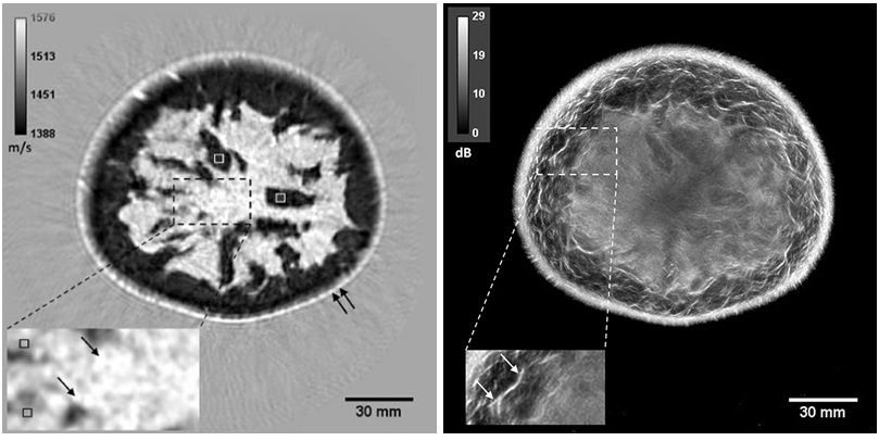
A novel AI tool aids articular cartilage segmentation (Photo courtesy of RSIP Vision)
The segmentation is carried out by classifying image pixels (or voxels, in 3D cases) using random forest classifiers, which delineate the boundaries between points in feature space belonging to different classes. The random forest is composed of an ensemble of decision trees, trained to assign membership value to either the lesion or the background group. To construct each tree, a different bootstrap subset of the training data is chosen at random. Since trees are uncorrelated, the overall decision in random forest is consistent by using a majority vote of trees with different structure.
“Our new segmentation tool makes it easier to pinpoint specific points and boundaries in images, which in turn leads to greater accuracy during surgeries without being dependent on the capability and experience of a specific individual,” said Ron Soferman, founder & CEO of RSIP Vision. “RSIP Vision will continue to drive innovation in image analysis across the medical verticals through custom software, advanced algorithm development and custom technologies.”
“Analyzing the parameters of the lesion and its boundaries allows the surgeon, along with the patient, to choose the ideal cartilage repair technique,” said orthopedic surgeon Shai Factor, MD, of Tel Aviv Sourasky Medical Center (Israel). “Additionally, in cases where cartilage transfer is the chosen option, this technology will make it possible to map the donor cartilage area as well and plan the surgery in the best way that will lead to better outcomes.”
Chondral lesions are prevalent among young and active patients, and due to the avascular nature of articular cartilage, healing potential is limited. In many cases, chondral lesions limit the athlete’s ability to participate in sports and even affect their daily activities. Cartilage segmentation is a crucial tool that aids the physician in choosing optimal treatment for the patient, including mosaicplasty, micro-fracture, osteochondral autograft transfer system (OATS), or autologous chondrocyte implantation.
Related Links:
RSIP Vision

Digital Color Doppler Ultrasound System
MS22Plus

Ultrasonic Pocket Doppler
SD1

Mobile X-Ray System
K4W

Pocket Fetal Doppler
CONTEC10C/CL