Innovative new Virtual Reality tool Under Development
By MedImaging International staff writers
Posted on 17 Jan 2017
Researchers are in the process of developing a tool that combines a virtual reality headset and anatomical 3D modeling of imaging data, to enable clinicians to navigate through the colon and search for polyps and other tumors.Posted on 17 Jan 2017
The tool is one several innovations resulting from several decades of research that is currently focused on developing software to help doctors, surgeons, and radiologists perform diagnoses and make decisions using 3D modeling, Virtual Reality (VR) and big data.
.")
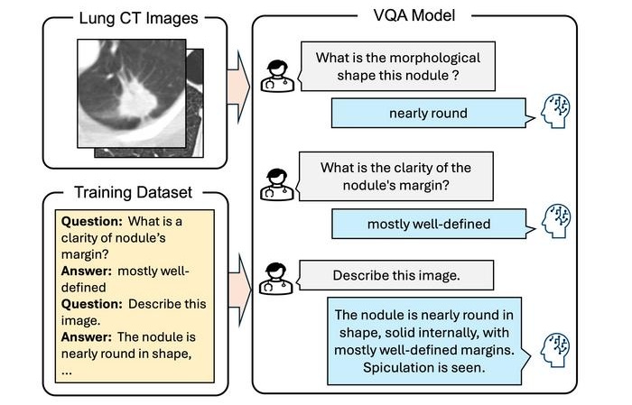
Image: The animation shows how researchers are using 3D virtual reality, and machine learning for diagnosis (Photo courtesy of GE Healthcare).
The researchers from the GE Healthcare Global Center of Excellence in Medical Imaging Software are located in Buc, France, and are using images from GE scanners for targeted 3D modeling of organs in the human body using virtual reality, and VR headsets.
The new technology enables a clinician to observe an organ or structure from all angles, in cross-sectional view, in 3D and in flat mode. In addition, a surgeon can combine this with the use of a 3D printer to be able to handle a replica of an organ before an operation. The researchers are using the Internet cloud to share images from 30,000 imaging scanners for their research that also includes machine-learning techniques.
Software Director, Jérôme Gonichon, at GE Healthcare, said, “This is an ongoing development process. Today, we are collecting data from the way that we are using our equipment so that we can improve it. Tomorrow, we can teach the machine to recognize cancer by itself.”

High-Precision QA Tool
DEXA Phantom

Digital Intelligent Ferromagnetic Detector
Digital Ferromagnetic Detector

X-Ray Illuminator
X-Ray Viewbox Illuminators

Ultrasonic Pocket Doppler
SD1