3-D Human Heart Printed Using Imaging Techniques
By MedImaging International staff writers
Posted on 05 Jul 2015
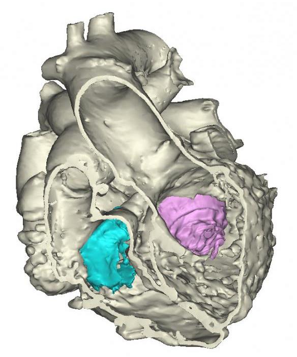
Congenital heart specialists have printed a 3-D anatomic model of a patient’s heart by integrating CT and Echocardiography techniques.Posted on 05 Jul 2015
This is the first time that researchers have integrated Computed Tomography (CT) and 3-D Transesophageal Echocardiography (3DTEE) to print a hybrid 3-D model of a heart. CT provided enhanced visualization of the heart’s external anatomy while 3DTEE provided visualization of valve anatomy. Magnetic Resonance Imaging (MRI) could also be integrated into the hybrid image to further enhance the 3-D model.

Image: 3-D Model of the Heart (Photo courtesy of Materialise).
The model was created by researchers at the Spectrum Health Helen DeVos Children’s Hospital (Grand Rapids, MI, USA). The researchers used the Mimics Innovation Suite software made by Materialise (Leuven, Belgium) to register images from the two imaging modalities, and selectively integrated datasets to create an anatomically accurate 3-D model of the heart. The model was printed using Materialise’s HeartPrint Flex technology.
Multiplanar reformatting enables virtual dissection of the heart, and helps visualize underlying pathology, such as heart defect. Hybrid 3-D models could also be used to help cardiologists plan trans-catheter or surgical interventions.
Joseph Vettukattil, MD, co-director and 3-D/4-D echocardiography researcher from the Helen DeVos Children’s Hospital, said, “This is a huge leap for individualized medicine in cardiology and congenital heart disease. The technology could be beneficial to cardiologists and surgeons. The model will promote better diagnostic capability and improved interventional and surgical planning, which will help determine whether a condition can be treated via transcatheter route or if it requires surgery.”
Related Links:
Helen DeVos Children’s Hospital
Materialise


