Automatic Processing Module Enhances Digital Imaging Workflow
By HospiMedica staff writers
Posted on 30 Jul 2008
A fully automatic processing module dramatically improves the digital imaging workflow while a "one click"-processing feature allows instant optimization of the digital image. Soft, standard, or sharp processing can easily be applied to satisfy the specific needs of the end user.Posted on 30 Jul 2008
With the latest version of image Clarity Enhancement processing software, developed by iCRco (Torrance, CA, USA), an equalized visualization of bone and soft tissue detail produced is excellent. "We have added a number of scientists that are very experienced in medical imaging to the iCRco team. The team has done a remarkable job with this software. The image quality that is now being produced by iCE 2 will not go unnoticed in this industry," stated Stephen Neushul, head engineer and president of iCRco, Inc.
The module can be added to any existing iCRco computed radiography (CR) device. The software enables the equalized display of enhanced details in bone structure and soft tissue at the same time. ICE 2 will advance the quality of patient care and improve the workflow of the imaging professional.
ICRco provides innovative digital X-ray imaging system, including a complete range of X-ray imaging hardware, picture archiving and communication systems (PACS), software, and integrated systems for the medical, dental, veterinary, and non-destructive testing markets.
Related Links:
ICRco



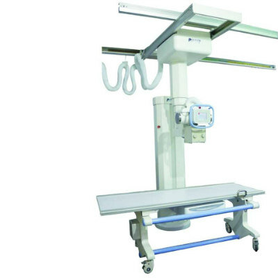

Digital Radiography System
DR-300
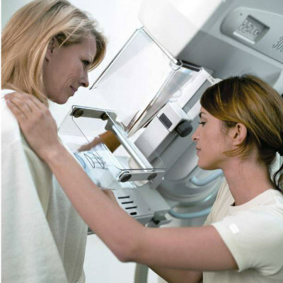

Mammo DR Retrofit Solution
DR Retrofit Mammography
Silver Member

X-Ray QA Device
Accu-Gold+ Touch Pro


Breast Localization System
MAMMOREP LOOP