Groundbreaking Technology to Enhance Precision in Emergency and Critical Care
Posted on 12 Mar 2026
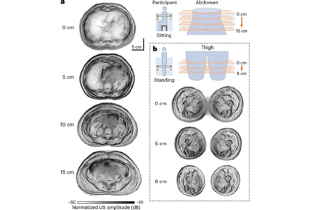
Rapid and accurate imaging is essential for diagnosing life-threatening conditions such as myocardial infarction, heart failure, and pulmonary embolism. However, conventional ultrasound imaging of the heart and lungs is often limited by rib bones, which reflect and scatter ultrasound waves and create blind spots in diagnostic images. Researchers have now developed a new technology designed to overcome this long-standing limitation and improve thoracic ultrasound imaging.
Scientists at The University of Hong Kong (HKU, Hong Kong, China) have developed SonoMeta, an artificial intelligence (AI)-powered ultrasound MetaLens that enables clearer imaging of organs located behind the rib cage. The system uses metamaterial-based microstructures that precisely control how ultrasound waves travel through the body, reducing reflection and scattering caused by rib bones.
")
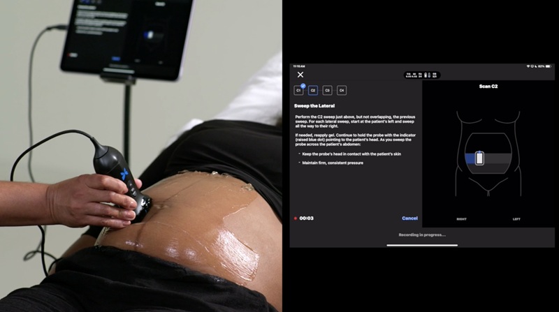
In conventional ultrasound examinations, clinicians must position the probe between the ribs to avoid bone interference. This limits imaging angles and may prevent full visualization of cardiac structures. In many cases, patients are referred for X-ray or CT scans to obtain clearer images, exposing them to radiation. The SonoMeta MetaLens modifies ultrasound wave propagation so that sound waves can pass through rib structures more effectively. This approach allows clinicians to visualize structures up to approximately 10 centimeters behind the rib cage, including heart valves and surrounding tissues.
Preclinical testing demonstrated improvements in imaging depth, clarity, and diagnostic accuracy compared with traditional ultrasound techniques. The technology could have significant value in emergency medicine, intensive care units, and ambulance settings where rapid imaging is critical. By enabling ultrasound scans directly through the rib cage, clinicians may be able to diagnose conditions such as heart failure, myocardial hypertrophy, valvular diseases, and pulmonary edema more quickly.
SonoMeta may also improve ultrasound-based screening of lung conditions. Because conventional ultrasound struggles to penetrate both air-filled lung tissue and ribs, early detection of lung abnormalities has been difficult. The new MetaLens allows clearer imaging of the thoracic cavity, potentially reducing reliance on radiation-based imaging such as CT scans. The research team has completed initial validation, simulation modellin,g and preclinical studies.
The developers are now working to refine the technology and create a portable MetaLens accessory that can be attached to existing ultrasound probes. If successfully implemented, the system could enable rapid, radiation-free thoracic imaging in bedside care, emergency settings, and telemedicine, expanding access to noninvasive cardiovascular and pulmonary diagnostics.
“Our goal is to leverage innovative engineering to address significant clinical challenges,” said Professor Nicholas X. Fang, who led the research team. “The ‘SonoMeta’ not only enhances image quality but, more importantly, makes diagnoses more precise, safe, and timely, truly embodying a patient-centered approach to healthcare.”
Related Links:
HKU