Motion Compatible Neuroimaging Device Enables Walking PET Brain Scans
Posted on 08 Aug 2024
Traditional Positron Emission Tomography (PET) scanners require patients to remain still during imaging. This is challenging for diagnosing conditions like Parkinson’s disease, where patients often experience involuntary movements. Such symptoms can make it more difficult to conduct scans when the condition advances, as conventional brain imaging demands complete immobility. Now, an upright neuroimaging device that allows patients to move around while undergoing a brain scan could help address these issues with traditional PET scanners.
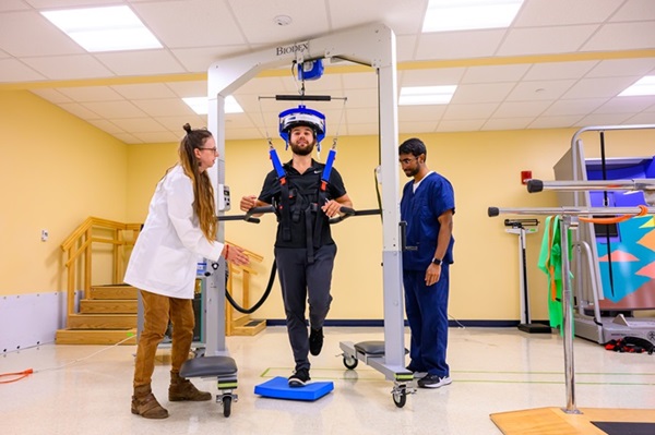
The prototype of the new device called an Ambulatory Motion-enabling PET, or AMPET, is an upgrade of an earlier scanner built by a team of neuroscientists, physicists and engineers at West Virginia University (WVU, Morgantown, WV, USA). AMPET is lighter and designed to be worn like a hard hat, providing balanced support on top, allowing it to move with the patient's head. This mobility allows patients to walk around while wearing the device. The team conducted real-world tests to evaluate the accuracy of AMPET and to identify areas for improvement. For these tests, outpatient volunteers who were already scheduled for standard scans and receiving imaging medications were enrolled. These volunteers wore the AMPET helmet and walked in place while the team monitored for motion tolerance and analyzed neural activity in the brain regions associated with movement.

The study successfully recorded brain activity in areas that coordinate leg movement as the patients walked, confirming the device’s capability. This outcome was further supported by scans from a patient with a prosthetic leg, showing significant brain activity in the region associated with his natural leg. The findings, reported in Nature Communications Medicine, highlight the potential of AMPET in clinical and research settings. Plans to enhance the device include adding motion tracking technology and expanding the helmet size to cover larger brain areas. AMPET could also benefit neuroscience research into natural human behaviors like gestures, conversation, and balance, and it has potential applications in treating PTSD, studying mindfulness meditation, and integrating with virtual reality technologies.
“What we demonstrated in the study is that when the patients walk, it’s not moving relative to the head and that’s what allowed us to get a relatively clean image,” said Julie Brefczynski-Lewis, research assistant professor in the Department of Neuroscience at the WVU School of Medicine. “To be able to image the brain in motion, we’re showing that there’s a whole new field that could open up because of our device.”
Related Links:
WVU


 Guided Devices.jpg)


