Non-Invasive Technique Combines Cardiac CT with AI-Powered Blood Flow for Heart Bypass Surgery
Posted on 08 Apr 2024
Researchers have tested a new approach to the guidance, planning, and conducting of heart bypass surgery on patients for the first time, demonstrating that non-invasive cardiac CT, with AI-powered blood flow scanning, is safe and feasible.
In the FAST TRACK CABG study, overseen by a research team at the University of Galway (Galway, Ireland; ), heart surgeons planned and carried out coronary artery bypass grafting (CABG). The procedure was performed based only on non-invasive cardiac-CT scan images using GE Healthcare’s (Chicago, IL, USA) Revolution CT, along with HeartFlow, Inc.’s (Redwood City, CA, USA) AI-powered blood flow analysis of the patient’s coronary arteries. This first-of-its-kind human study demonstrated 99.1% feasibility, indicating that heart bypass surgery performed without utilizing invasive diagnostic catheterization is feasible and safe, based on good diagnostic accuracy provided by the cardiac CT scan and AI-powered blood flow analysis. The study found similar outcomes in terms of safety and effectiveness in patients who had previously undergone bypass surgery guided by conventional invasive angiography, which requires the insertion of a catheter through an artery in the wrist or groin to access diseased arteries and the use of dye to visualize blockages.

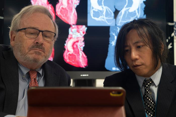
Conducted across leading cardiac care hospitals in Europe and the US, the study included 114 patients with severe coronary artery blockages, impacting their heart blood flow. The cardiac CT scans provided in the study delivered exceptional resolution, producing images on par with, or superior to, those obtained through invasive contrast dye injections directly into the heart’s arteries through a catheter. Throughout the trial, detailed cardiovascular imagery and data analyses were performed by the University of Galway team and shared via telemedicine with surgeons at participating hospitals. The HeartFlow Analysis performs an AI-powered blood flow analysis called Fractional Flow Reserve derived from CT (FFRCT) to quantify how poorly the narrowed vessel provides blood to the heart muscle, assisting the surgeon in identifying which of the patient’s vessels require a bypass graft. The FAST TRACK CABG trial's findings indicate that this innovative, less invasive approach to heart bypass surgery is as safe and effective as traditional methods. The study highlights the potential for replacing the risks associated with invasive procedures with non-invasive cardiac CT imaging and AI-based blood flow analysis, offering a significant advancement in cardiac care and surgery planning.
“The results of this trial have the potential to simplify the planning for patients undergoing heart bypass surgery,” said trial chairman Professor Patrick W Serruys, Established Professor of Interventional Medicine and Innovation at University of Galway. “The potential for surgeons to address even the most intricate cases of coronary artery disease using only a non-invasive CT scan, and FFRCT represents a monumental shift in healthcare. Following the example of the surgeon, interventional cardiologists could similarly consider circumventing traditional invasive cineangiography and instead rely solely on CT scans for procedural planning. This approach not only alleviates the diagnostic burden in cath labs but also paves the way for transforming them into dedicated ‘interventional suites’- ultimately enhancing patient workflows.”
Related Links:
University of Galway
GE Healthcare
HeartFlow, Inc.


