New Cost-Effective PET Scanning Technology Improves Image Resolution for Whole-Body Scans
Posted on 30 Jun 2023
Whole-body PET/CT imaging is widely utilized for cancer staging, restaging, and assessing patient responses to treatments. However, its diagnostic accuracy can be compromised when dealing with very small lesions or weak signals. Now, a novel technology, dubbed "Augmented Whole-Body Scanning via Magnifying PET" (AWSM-PET), has been shown to enhance the image resolution and system sensitivity of clinical whole-body PET/CT imaging. This cost-efficient technology employs high-resolution add-on detectors that simultaneously scan a patient during a standard whole-body PET scan.
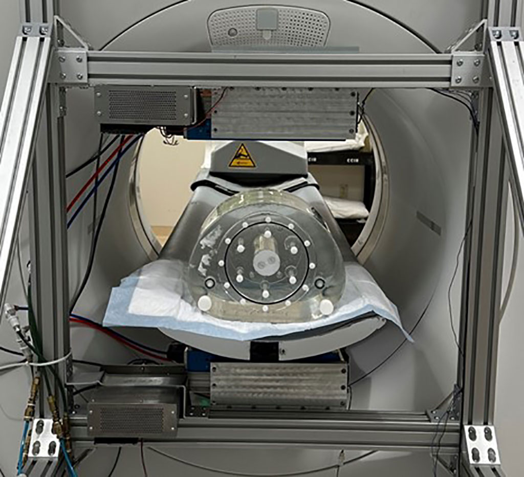
The AWSM-PET technology developed by researchers at Washington University (St. Louis, MO, USA) utilizes two high-resolution PET detectors placed outside the scanner's axial imaging field of view as an "outsert" device. Each outsert panel contains 32 LSO crystal arrays, each with 30x30 elements (0.97x0.97x10.0 mm each). The device acquires high-resolution PET data simultaneously as a patient undergoes a whole-body PET scan, without requiring any additional scanning time. Additionally, the researchers have developed custom reconstruction and correction algorithms to jointly reconstruct the data.

The researchers tested their technology using a prototype AWSM-PET device on a Siemens Biograph Vision PET/CT scanner. They imaged cylindrical phantoms with varying-sized tumor inserts and noted a significant improvement in image resolution when data from the outsert device were incorporated. The outsert detectors demonstrated superior spatial, energy, and timing resolution, with a system-level coincidence resolving time of 217 ps – a time-of-flight application suitability. The researchers are planning to initiate a pilot human imaging trial later this year to compare the diagnostic accuracy of AWSM-PET with the standard whole-body PET/CT.
“Our novel AWSM-PET prototype helps to tackle two of the key limitations in whole-body PET imaging: image resolution and overall system sensitivity,” said Yuan-Chuan Tai, PhD, associate professor of radiology at Washington University. “The additional high-resolution data from the AWSM-PET device can enhance the overall image resolution and reduce statistical noise. The potential improvement in diagnostic accuracy of clinical whole-body PET/CT may benefit cancer patients.”
Related Links:
Washington University


