Novel Imaging Agent Non-Invasively Identifies Early Stage Pulmonary Fibrosis
Posted on 22 Jun 2023
Pulmonary fibrosis, a lethal condition, typically presents a survival rate of three to five years post-diagnosis. Diagnosing the disease in its advanced stages is relatively easy, yet pinpointing the earliest stages, when treatments are most effective, can prove challenging. At present, the standard clinical diagnosis of pulmonary fibrosis depends on breath tests and CT scans to observe changes in lung structure. However, these anatomical details often fall short of detecting the early indicators of the disease. Now, a newly-formulated PET imaging agent offers a non-invasive means to spot pulmonary fibrosis in its nascent stages, reducing unnecessary biopsies and facilitating earlier treatment initiation.
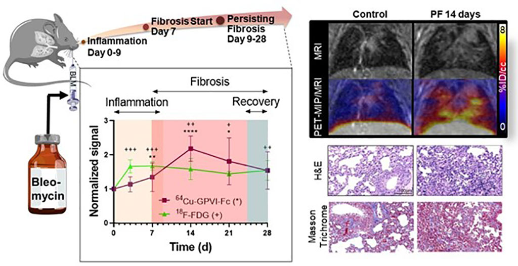
Pulmonary fibrosis in patients results in lung tissue remodeling due to the increased deposition of extracellular matrix fibers such as collagen I-III, fibronectin, and fibrinogen. In a study, researchers at Eberhard Karls University of Tübingen (Tübingen, Germany) used an imaging agent named 64Cu-GPVI-Fc, designed to target these extracellular matrix fibers, in order to detect pulmonary fibrosis in a mouse model. The results were then compared to histological findings and 18F-FDG PET imaging results. The researchers found that 64Cu-GPVI-Fc demonstrated substantial uptake in lungs afflicted with pulmonary fibrosis, which was in line with the histological outcomes. Unlike the 18F-FGD PET imaging results, the uptake of 64Cu-GPVI-Fc was exclusively associated with pulmonary fibrosis activity in the lung tissues and did not detect any inflammation.

“In a disease with such a large impact on the patients’ quality of life and with such a reduced life expectancy after diagnosis, it is critical that proper diagnosis and treatment follow-up methods are specific and sensitive enough that optimal medical care can be given. We believe 64Cu-GPVI-Fc takes us one step closer to personalized medicine for pulmonary fibrosis,” said Nicolas Bézière, PhD, head of Imaging of Infection and Inflammation at the Werner Siemens Imaging Center, Department of Preclinical Imaging and Radiopharmacy at Eberhard Karls University of Tübingen. “We hope that this approach based on a tracer targeting a range of extracellular matrix fibers will provide a new way to view the ‘complete picture’ of pulmonary fibrosis progression and act as a new method to monitor treatment efficacy. Furthermore, fibrosis is not limited to the lungs, it can develop in other organs and lead to a loss of their function. Thus, we can foresee the transfer of this approach to other fibrotic diseases.”
Related Links:
Eberhard Karls University of Tübingen


