AI Helps Optimize CT Scan X-Ray Radiation Dose
Posted on 10 Mar 2023
Computed tomography (CT) is a highly effective and extensive diagnostic tool utilized by modern medicine. Unfortunately, there is growing concern regarding the increasing number of patients undergoing CT scans, and the considerable amount of X-ray radiation to which they are exposed. The ALARA principle, commonly known as "As Low As Reasonably Achievable," implies that a patient should receive the most significant diagnostic benefit with minimal radiation exposure. In practical terms, this principle requires a trade-off since decreasing the level of radiation administered typically results in poorer CT image quality. Accordingly, medical professionals must strike a balance between obtaining high-quality CT images and minimizing a patient's exposure to X-rays to reduce the risk of misdiagnosis.
To strike a balance between image quality and radiation exposure during CT scans, healthcare professionals, including radiologists, may employ an optimization strategy. First, they observe actual images generated by the tomographer to identify abnormalities such as tumors or unusual tissue. Statistical methods are then utilized to calculate the optimal radiation dose and tomographer configuration. This procedure can be generalized by adopting reference CT images obtained by scanning specially designed phantoms that comprise inserts of varying sizes and contrasts, which represent standardized abnormalities. However, manual image analysis is incredibly time-consuming. To address this issue, a team of researchers from the University of Florence (Florence, Italy), in collaboration with radiologists and medical physicists, examined if this process could be automated by using artificial intelligence (AI). The team created and trained an algorithm - a “model observer” - based on convolutional neural networks (CNNs), which could analyze the standardized abnormalities in CT images as efficiently as a professional.

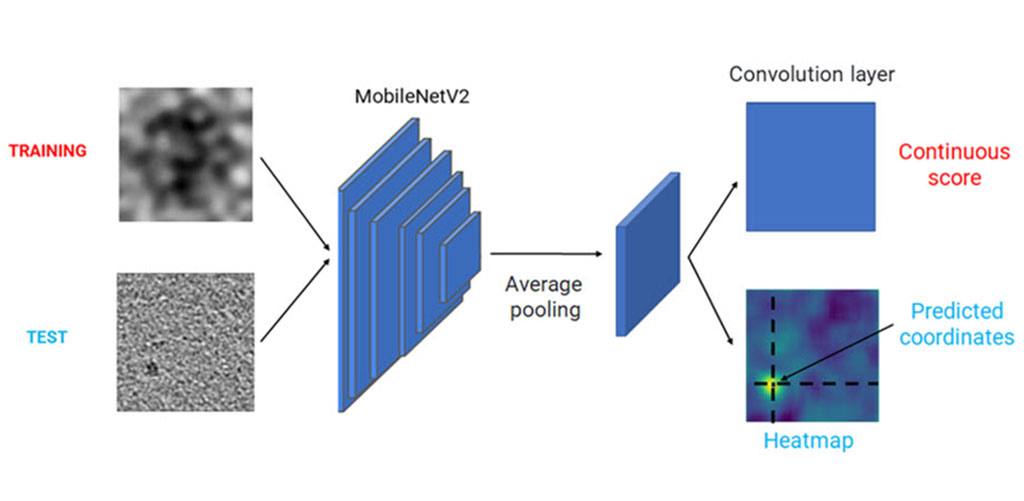
The team needed sufficient training and testing data for their model, for which 30 healthcare professionals visually examined 1000 CT images in a phantom mimicking human tissue. The phantom contained cylindrical inserts of varying diameters and contrasts, and the observers had to identify whether an object was present in the image as well as indicate the level of confidence in their assessment. This generated a dataset of 30,000 labeled CT images captured using various tomographic reconstruction configurations, accurately reflecting human interpretation. The team then implemented two AI models based on different architectures - UNet and MobileNetV2 – and modified the base design of these architectures to allow them perform both classification ("Is there an unusual object in the CT image?") and localization ("Where is the unusual object?"). The models were then trained and tested using images from the dataset.
The research team conducted statistical analyses to evaluate various performance metrics to ensure that the model observers accurately emulated how a human would assess CT images of the phantom. The researchers are optimistic that with further efforts, their model can become a viable mechanism for automated CT image quality assessment. They are confident that applying their AI model observers on a larger scale will enable faster and safer CT evaluations than ever before.
“Our results were very promising, as both trained models performed remarkably well and achieved an absolute percentage error of less than 5%,” said Dr. Sandra Doria of the Physics Department at the University of Florence who led the research team. “This indicated that the models could identify the object inserted in the phantom with similar accuracy and confidence as a human professional, for almost all reconstruction configurations and abnormalities sizes and contrasts.”
Related Links:
University of Florence


