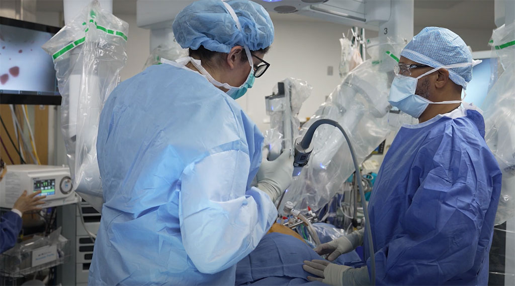
Same-Day Procedure to Diagnose and Treat Lung Cancer Combines CT Scan and Robotic Video-Assisted Thoracic Surgery
Posted on 22 Feb 2022
A new approach combines the use of a robot for minimally invasive bronchoscopy for early lung cancer detection with same-day surgery to remove lung tumor.
A team of pulmonary and thoracic experts at MedStar Georgetown University Hospital (Washington, DC, USA) has adoped this new approach for lung cancer diagnosis and surgical removal of the tumor on the same day, thereby increasing the opportunity for limited follow-up therapy – or even curing the cancer. Previously this process of diagnosis and treatment could take several weeks - a critical time when cancer can continue to grow, and patients can experience increased anxiety about their health and prognosis.

In this new approach – Combined Robotic Assisted Thoracic Surgery (CRATS) – high-risk lung cancer patients first undergo a low dose CT scan of the lung to monitor for tumors. If a tumor is detected, the Ion robot bronchoscope is used to biopsy the lung tumor. If the lung tumor is found – through real-time biopsy – to be cancer, then the endobronchial ultrasound is used to determine if the cancer has spread to lymph nodes. If the lymph nodes are normal, then the patient undergoes surgery to remove the portion of the lung with the cancerous tumor using the DaVinci robotic video-assisted thoracic surgery system during the same anesthesia procedure. This new process – diagnosing lung cancer and removing the tumor at the same time - saves valuable time by removing the cancer right when it is detected, thereby limiting the opportunity for spread of disease and allowing for the start of additional therapies sooner if needed. In some instances, this approach can be curative of the cancer.
The Ion robot is a “game-changing” tool that enables surgeons to reach peripheral nodules in the lung to diagnose lung cancer sooner, thereby increasing the opportunity for earlier diagnosis and treatment of lung cancer, the leading cause of cancer deaths worldwide for both men and women. Ion endoluminal is a minimally invasive robotic-assisted system that helps address a challenging aspect of lung biopsy by enabling physicians to obtain tissue samples from deep within the lung. While survival rates for lung cancer have improved over the past decade in part due to early screening, physicians know that improved outcomes are directly linked to early detection. Coupled with regular screening for those at high risk, the Ion system makes possible earlier and more accurate detection of cancer, especially for small nodules.
In addition to giving surgeons real-time guidance for improved accuracy, the Ion robot can navigate through small and tortuous airways to reach nodules in any airway segment within the lung with its flexible biopsy needle that can pass through very tight bends to collect tissue in the peripheral lung. The catheter’s 2 mm working channel can also accommodate other biopsy tools, such as biopsy forceps and cytology brushes, if necessary. The system’s fiber optic shape sensor technology, developed by Intuitive, is to provide the physician with the precise location and shape information of the catheter throughout the navigation and biopsy process.
“Coupling this early diagnosis with same-day surgery for lung cancer is a win for patients,” said Eric D. Anderson, MD, director of Interventional Pulmonology at MedStar Georgetown University Hospital. “We know that the earlier we find the nodule in the lung and the smaller it is, that has the greatest impact on mortality. If we find the nodule at 1 mm, there is 90% chance of survival. At 2 mm, there is 80%, and at 3 mm 70%. Combine those odds with the potential immediate removal of the tumor, and the Ion robot gives patients a real chance for better outcomes.”
Related Links:
MedStar Georgetown University Hospital


