New Imaging Technology Detects Gastric Lymphomas
By MedImaging International staff writers
Posted on 13 Oct 2021
A new imaging technique for the detection of gastric mucosa-associated lymphoid tissue (MALT) lymphomas could reduce invasive gastroscopy biopsies, according to a new study.Posted on 13 Oct 2021
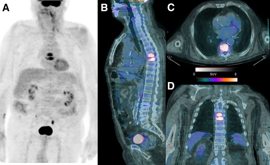
The study, by researchers at Memorial Sloan-Kettering Cancer Center (MSKCC; New York, NY, USA), the Medical University of Vienna (MedUni; Austria), and other institutions, explored if [68Ga]Pentixafor, a positron emission tomography (PET) tracer that targets the chemokine receptor CXCR4 (which is overexpressed in MALT lymphoma), could serve as a potential alternative to follow-up biopsies for assessment of residual disease following first-line H. pylori eradication.
")
Image: MALT lymphomas express the chemokine receptor CXCR4 (Photo courtesy of MedUni)
The study, which included [68Ga]Pentixafor-PET and magnetic resonance imaging (MRI) scans in 26 gastric MALT lymphoma patients and 20 control group patients without lymphoma, showed that accuracy, sensitivity, specificity, and positive and negative predictive value of [68Ga]Pentixafor-PET for detection of residual gastric MALT lymphoma at follow-up were 97%, 95%, 100%, 100%, and 92.9%, respectively. Maximum and mean PET standardized uptake values also showed correlation with immunohistochemistry-based CXCR4 cell counts. The study was published on September 15, 2021, in Blood.
“Post-treatment evaluation of gastric MALT currently relies on esophagogastroduodenoscopy with histological assessment of biopsies,” concluded lead author Marius Mayerhöfer, PhD, of the MedUni department of biomedical imaging and image-guided therapy, and colleagues. “If a sufficiently high CXCR4 expression is detected at the initial diagnosis of MALT lymphoma, the new imaging could replace repeated gastroscopies in the course of the disease in the future, or at least increase the time intervals between gastroscopies.”
CXCR4 is an alpha-chemokine receptor specific for stromal-derived-factor-1 (SDF-1), a molecule with potent chemotactic activity for lymphocytes. After attachment to SDF-1, the CXCR4 protein activates signaling pathways inside the cell that help regulate cell growth and proliferation. CXCR4 is also involved in cell migration cells, especially hematopoietic stem cells in the bone marrow.
Related Links:
Memorial Sloan-Kettering Cancer Center
Medical University of Vienna

Mammo DR Retrofit Solution
DR Retrofit Mammography

Ultrasonic Pocket Doppler
SD1

X-ray Diagnostic System
FDX Visionary-A

Biopsy Software
Affirm® Contrast