Organ-Based PET Scanner Improves Image Quality
By MedImaging International staff writers
Posted on 07 Oct 2021
An organ-based positron emission tomography (PET) camera employs detectors based on silicon photomultipliers rather than traditional instrumentation.Posted on 07 Oct 2021
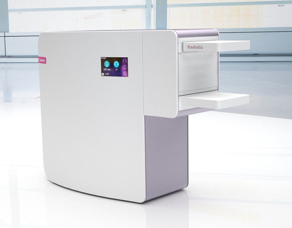
The Radialis Medical (Thunder Bay, Canada) camera is a targeted PET imaging system that uses state-of-the-art silicon photomultiplier data acquisition sensors (combining high signal gain and low signal noise) positioned in close proximity to the organ of interest for a higher-quality image with a smaller field of view. By using a patented seamless detector array that can be rotated, and by positioning patients so that the targeted organ is within the field of view, full coverage of the target area, without gaps or dead zones, can be achieved.

Image: The Radialis organ-targeted PET camera (Photo courtesy of Radialis Medical)
For example, during breast imaging, the slim detector heads enable tight positioning against the chest wall. The PET camera's detector electronics also include integrated cooling to enhance signal quality and high-throughput data acquisition that has been optimized for the low-dose imaging the camera is capable of due to high radiotracer sensitivity. Peak absolute sensitivity at the center of the field of view is 3.5%, with normalized total absolute sensitivity of 2.4%. The high sensitivity translates into an in-plane image resolution of up to 2.5mm.
“Compared to whole-body PET scanners, an organ-targeted PET camera positions detectors in close proximity to the organ of interest for a higher quality image of a smaller field of view,” said Michael Waterston, CEO of Radialis Medical. “The device would be an ideal addition to clinics with whole-body PET systems due to its improved sensitivity, resolution, and flexibility that enables precision imaging of multiple organs with as little as 1/10th of the normal radiotracer dose.”
PET is a nuclear medicine imaging technique that produces a 3D image of functional processes in the body. The system detects pairs of gamma rays emitted indirectly by a positron-emitting radionuclide tracer, such as 21-[18F]fluorofuranylnorprogesterone (FFNP), a progestin analog. Computer analysis is then used to display tracer concentrations within the body.
Related Links:
Radialis Medical


