DEXA Predicts Diabetes in Obese Menopausal Women
By MedImaging International staff writers
Posted on 13 Sep 2021
Dual-energy x-ray absorptiometry (DEXA) beats traditional clinical obesity measures for predicting diabetes and heart disease in older women, according to a new study.Posted on 13 Sep 2021
Researchers at Stanford University (CA, USA), the University of Illinois in Chicago (UIC; USA), and other institutions analyzed data on 9,744 postmenopausal women (50-79 years of age) who underwent a DEXA scan and were free of cardiovascular disease (CVD) and diabetes at baseline. The aim of the study was to see if DEXA estimates of adiposity in older women could improve risk prediction for cardiometabolic diseases better than the traditional surrogates - body mass index (BMI), waist circumference (WC), and waist-to-hip ratio (WHR).

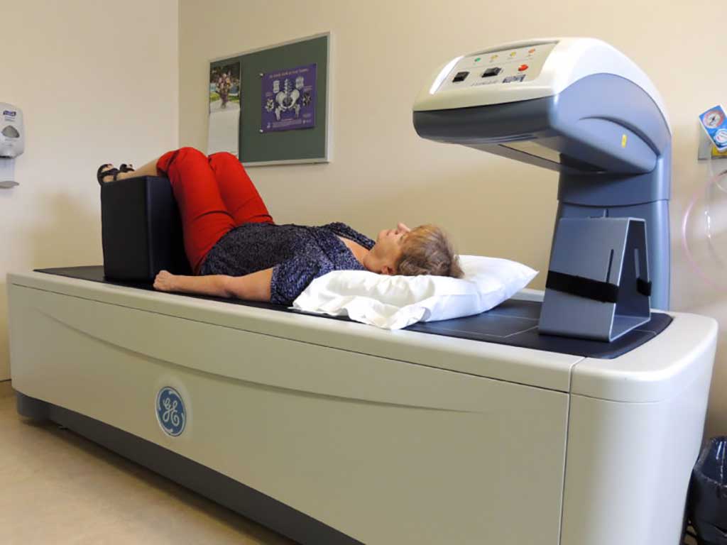
Image: Bone density measurements can also help diagnose diabetes in older women (Photo courtesy of Alamy)
The results revealed a total of 1,327 diabetes, 1,266 atherosclerotic CVD, and 292 heart failure (HF) cases, as well as 1,811 deaths from any cause accrued during a median follow-up of 17.2 years. The largest hazard ratio per adiposity measure for diabetes and atherosclerotic CVD was percentage of trunk fat (%TrF), which was also the only adiposity measure to demonstrate statistically significant improved concordance probability estimates over BMI, WC, and WHR. The study was published on August 31, 2021, in Mayo Clinic Proceedings.
“The single most striking finding of our multivariable analyses was the magnitude of association observed between %TrF and the risk of incident diabetes,” concluded lead author Deepika Laddu, PhD, of the University of Chicago. “Using DEXA to improve risk prediction for cardiometabolic diseases among patients already being screened for osteoporosis presumably would have little impact on cost of care, and has the potential to impact public health substantially.”
DEXA is a means of measuring bone mineral density (BMD) using spectral imaging. Two X-ray beams, with different energy levels, are aimed at the bones. When soft tissue absorption is subtracted, BMD can be determined from the absorption of each beam. DEXA is most often used to diagnose and monitor osteoporosis.
Related Links:
Stanford University
University of Illinois in Chicago


