Nanowire Single-Photon Sensor Detects Cerebral Blood Flow
By MedImaging International staff writers
Posted on 30 Aug 2021
Superconducting nanowire single-photon detectors (SNSPDs) could enable precise measurement of cerebral blood flow, according to a new study.Posted on 30 Aug 2021
Adapted for diffuse correlation spectroscopy (DCS) imaging use by researchers at Massachusetts General Hospital (MGH; Boston, USA) and the Massachusetts Institute of Technology (MIT; Cambridge, MA, USA), the SNSPDs sensors consist of a thin film of superconducting material with excellent single-photon sensitivity and detection efficiency. While commonly used in optical quantum information, telecommunications, and space communications, SNSPDs were seldom used in biomedicine, till now.

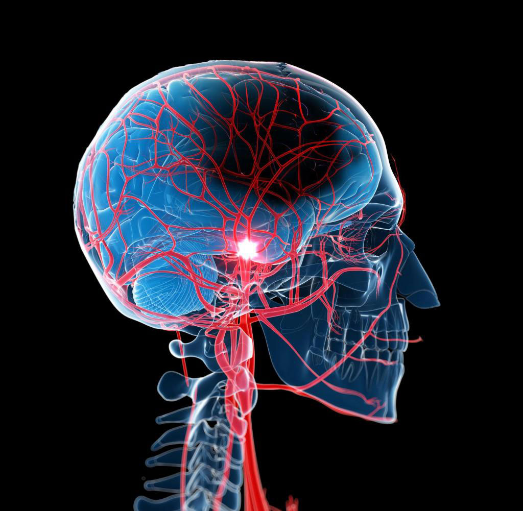
Image: Novel nanowire sensors detect cerebral blood flow non-invasively (Photo courtesy of Getty Images)
To test the efficacy of the system, the researchers conducted cerebral blood flow measurements on 11 participants using a SNSPD-DCS system and a conventional single-photon avalanche photodiode (SPAD)-DCS system, both provided by Quantum Opus (Novi, MI,USA) . The SNSPD-DCS system operated at a wavelength of 1064 nm with two SNSPD detectors, whereas the quadruple SPAD-DCS system operated at 850 nm. The results showed that the SNSPDs outperformed SPADs in multiple parameters, such as time resolution, photon efficiency, and range of wavelength sensitivity.
The SNSPD-based DCS system showed significant improvement in signal-to-noise ratio (SNR) over SPAD-based DCS, as they received seven to eight times more photons than SPAD detectors, and at a double the efficiency. The 16 times increase in SNR allowed signal acquisition at 20 Hz at the same source–detector (SD) separation, allowing clear detection of arterial pulses. In addition, SNSPD-DCS was more effective in measuring cerebral blood flow during breath-holding and hyperventilation, and were in agreement with those obtained from PET and MRI studies. The study was published on August 19, 2021, in Neurophotonics.
“The SNSPD-DCS system facilitates higher photon collection, larger SD separations, and higher acquisition rates, leading to better accuracy,” concluded lead author Nisan Ozana, PhD, of MGH, and colleagues. “Given these advantages, this novel system may allow for a non-invasive and more precise measurement of cerebral blood flow, an important marker of cerebrovascular function, for adult clinical applications.”
DCS involves the illumination of tissues with near-infrared (nIR) lasers. The light is scattered by the movement of red blood cells (RBCs), and the diffraction pattern formed is analyzed to determine blood flow.
Related Links:
Massachusetts General Hospital
Massachusetts Institute of Technology
Quantum Opus


