Imaging Significantly Underestimates the Size of DCIS Tumors
By MedImaging International staff writers
Posted on 15 Apr 2021
A new study suggests that pre-operative imaging of high-volume ductal carcinoma in situ (DCIS) lesion size is inaccurate. Posted on 15 Apr 2021
Researchers at the University of British Columbia (UBC; Vancouver, Canada), Western University (UWO; London, Canada), and Mount Saint Joseph Hospital (Vancouver, Canada) undertook a retrospective review of clinicopathologic data of patients treated for DCIS with breast conserving surgery (BCS) between 2012 and 2018. Mammographic and sonographic lesion sizes were compared with final pathology sizes.

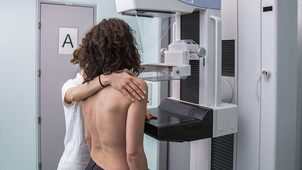
Image: Imaging can underestimate the actual size of a breast tumor (Photo courtesy of Getty Images)
The results showed that for 152 lesions visible on mammography, mean size on imaging was significantly smaller when compared to final pathology (2.3 centimeters versus 3.6 centimeters, respectively); the mean difference of 1.3 cm was a significant underestimation, with a correlation coefficient of 0.367. For the 48 lesions visible on ultrasound, the radiologic size was significantly smaller than pathologic size (1.7 centimeters versus 4.1 centimeters, respectively), but degree of underestimation was not significantly correlated. The study was published on March 23, 2021, in The American Journal of Surgery.
“Imaging size of DCIS tumors assessed by both mammography and ultrasound were significantly smaller when compared with their final size on pathology,” concluded lead author Rachel Liu, MD, of UWO, and colleagues. “Increase in the degree of underestimation by mammography was noted as the histological tumor size increased. This must be taken into consideration during surgical planning for maximizing the success of breast conserving surgery and minimizing return to the operating room.”
DCIS is the most common type of non-invasive breast cancer, in which the abnormal cells are contained inside the milk ducts. If DCIS is not treated, it may eventually develop into invasive breast cancer, which can spread outside the ducts into the breast tissue and then possibly to other parts of the body. Since DCIS cannot usually be felt as a breast lump or other breast change, most cases are diagnosed following routine screening with mammograms or ultrasound, appearing as micro-calcifications.
Related Links:
University of British Columbia
Western University
Mount Saint Joseph Hospital


