Novel Cardiac SPECT System Enhances Patient Comfort
By MedImaging International staff writers
Posted on 22 Oct 2020
An updated single-photon emission computed tomography (SPECT) scanner offers nuclear cardiology providers high level of image quality.Posted on 22 Oct 2020
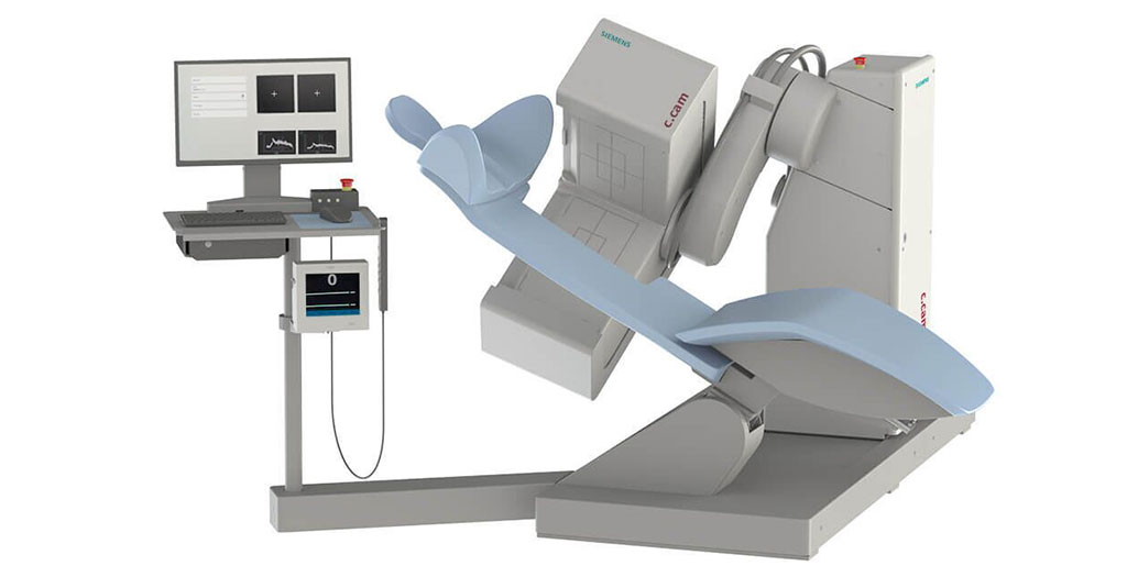
The Siemens Healthineers (Erlangen, Germany) c.cam Cardiac SPECT System features an integrated, bi-directional camera arm that permits easier patient positioning, and a comfortable, reclining patient chair with a 205 kilogram weight limit. The chair improves access to care by allowing the patient to remain seated at ease during scanning, minimizing respiratory motion and cardiac image artifacts. The chair does not contain pallet material, resulting in zero attenuation in focus area.

Image: The c.cam Cardiac SPECT System (Photo courtesy of Siemens Healthineers)
The camera is fixed at 90º to the reclining chair, creating a minimal patient-to-detector distance. Arm rotation and detector swivel motors deliver a circular SPECT motion, creating a small field of view optimized for cardiac procedures. Lightweight collimators and advanced digital detectors with five channel row-and-column decoding per individual photomultiplier tube (PMT) offers high image quality. The Windows-based system also contains a cybersecurity package that provides enhanced features to limit access by non-authorized personnel.
“This new version of the c.cam SPECT scanner is designed to provide an affordable, easy-to-install system that doesn’t sacrifice image quality and enhances the overall patient experience,” said John Khoury, vice president of molecular imaging at Siemens Healthineers North America. “With a small footprint, the c.cam can be installed in just two days with minimal room modeling requirements. Applications training can be completed in only three days.”
SPECT myocardial perfusion imaging (MPI) is a non-invasive imaging technique to evaluate the presence and severity of coronary artery disease (CAD) by detecting flow-limiting disease, risk-stratifying patients, and assessing and quantifying patient risk. It is most frequently used for perioperative cardiovascular evaluation and chest pain. Recent studies have shown that noninvasive SPECT scans of cardiac vessels are far better at spotting CAD than commonly prescribed exercise stress tests.


